. 2009 Jan;174(1):239-47.
doi: 10.2353/ajpath.2009.080627. Epub 2008 Dec 18.

A physical mechanism for coupling bone resorption and formation in adult human bone

Affiliations

A physical mechanism for coupling bone resorption and formation in adult human bone

Thomas Levin Andersen et al. Am J Pathol. 2009 Jan.

Abstract

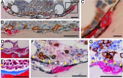
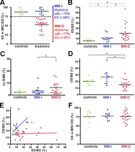
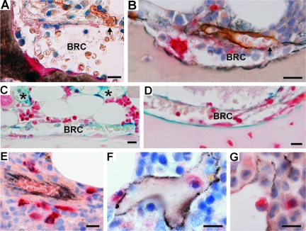
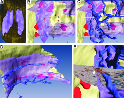
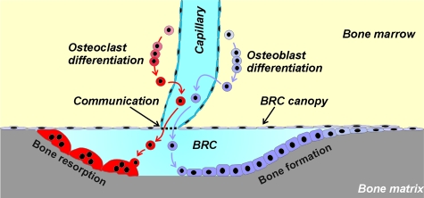
During skeletal remodeling, pre-osteoclasts and pre-osteoblasts are targeted to critical sites of the bone to resorb and reconstruct bone matrix, respectively. Coordination of site-specific recruitment of these two cell types is a prerequisite to maintain the specific architecture of each bone within strict limits throughout adult life. Here, we determined that the bone marrow microanatomy adjacent to remodeling areas is a central player in this process. By using histomorphometry and multiple immunostainings, we demonstrated in biopsies exhibiting coupled bone resorption and formation that osteoclasts and osteoblasts on the bone surface were always covered by a canopy of flat cells expressing osteoblast markers. In contrast, in biopsies in which this canopy was disrupted, bone formation was deficient. Three-dimensional visualizations revealed that this canopy covered the entire remodeling site and was associated with capillaries, thereby forming a previously unrecognized microanatomical entity. Furthermore, pre-osteoclasts were positioned along these capillaries. These findings led to a model that implicates vasculature in the site-specific recruitment of osteoclasts and osteoblasts and embraces the current knowledge on the molecular mechanism of bone remodeling.


Figures

Figure 1
Figure 1
The histological appearance of the BRC. A: Cross section of a complete BRC. The wall of the BRC is made of NCAM-positive (black) elongated cells lining the bone marrow. The bone-lining cells are also NCAM-positive. B: Another cross section of a BRC showing PIIINP immunoreactivity (black) in the elongated cells of the wall and in the adjacent CD34-positive capillaries (brown). C: High magnification of a TRAcP+ (red) OC, positioned at the periphery of the BRC (inset in A) and between the bone surface and an elongated NCAM-positive cell of the BRC canopy. D and E: The morphological appearance of BRCs as revealed by Masson’s trichrome, containing eroded (D) or osteoid (E) bone surfaces. The yellow arrows point to the BRC canopy. F and G: Two examples of disrupted BRC canopies. The canopy is not detected in the area highlighted by the white arrowheads, but is seen along the yellow arrows. Scale bars: 25 μm (C); 50 μm (A, B, D–G). BRC: bone remodeling compartment.
Figure 2
Figure 2
Correlation between the presence of intact BRC canopies and coupled bone erosion/formation. A: The proportions of erosion surfaces (ESs) in and out of BRCs were measured in bone biopsies from 9 control and 32 myeloma patients. Note that all control bones (green) show more than 75% ES in intact BRCs, whereas myeloma samples show values both above (blue) and less than 75% below (red). Therefore our analysis considered separately these two myeloma subpopulations, indicated as MM-I(ntact) and MM-D(isrupted), respectively. B–D: In the biopsies of each of these two MM groups and of controls, the extent of eroded surfaces (ES) (B), OC surfaces (Oc.S) (C), and osteoid surfaces (OS) (D) was measured and related to the total bone surface (BS). Note that the MM-D population shows the largest ES (B) and the smallest OS (D). E: Plotting osteoid surface (OS) versus eroded surface (ES) for each biopsy shows coupling between these two events in both the control and MM-I biopsies and uncoupling in the MM-D biopsies. F: The proportion of osteoid surfaces (OS) in and out of BRCs was measured in biopsies from each of the three groups. Note that all show the same average of ∼77% OS in intact BRCs. A: This is in contrast with erosion that may occur outside of intact BRCs. The statistical significances were calculated by using the Mann-Whitney test: *P < 0.05.
Figure 3
Figure 3
Histology revealing the physical relation between the BRC canopy and capillaries, and between capillaries and OC progenitors. A and B: Two histological sections passing through a contact point (arrows) between a CD34+ capillary (brown) and a BRC canopy. A: Erythrocytes can be clearly distinguished in the lumen of the capillary and inside the BRC. C and D: Ferritin was detected in the capillaries (asterisk) and in the BRCs of rabbits 5 minutes after intravenous injection, as visualized with Perls’ Prussian blue. E–G: TRAcP+ pre-OCs (red) were observed around (E) and in rare occasions within (F, G) CD34+ capillaries (black) of the bone marrow. Scale bars = 25 μm.
Figure 4
Figure 4
Three-dimensional reconstructions of BRCs and related microanatomical structures. A: Top view of two nearby BRCs allowing appreciation of the continuity of the BRC canopy (purple) and its sealing with the bone-lining cells (brown), thereby forming a closed compartment. B: Semilateral view of a BRC shown open to visualize the space comprised between the bone surface (pale yellow) bearing large mature OCs (red) and the BRC canopy (transparent purple). Small pre-OCs (orange) are visible at the bone marrow side of the BRC canopy C: The same BRC as in B, shown with the capillary network (blue). Note the position of pre-OCs along the capillaries. D: Another BRC. Large OCs (red) are visible on the bone surface through the BRC canopy (transparent purple). Note the correlation between the positions of these OCs and those of the capillaries at the bone marrow side of the BRC wall. E: Focus on the area framed in C, with superimposed histological picture (see Figure 3A), to illustrate the connection point of a capillary with the BRC canopy (arrow). The three-dimensional information contained in A–E can best be appreciated in the supplemental movies available at http://ajp.amjpathol.org. Supplemental Figure S2 at http://ajp.amjpathol. org also shows a low-magnification view of a piece of bone with BRCs and related (pre-) OCs, indicating the in situ position of the BRCs shown in A and B.
Figure 5
Figure 5
Model for coordinating site-specific recruitment of OC and OB progenitors to bone remodeling sites. The model summarizes the main features recognized in our study (see text). A bone remodeling site involves joint OC and OB activities on the bone surface [bone remodeling unit (BRU) or basic multicellular unit (BMU)2]. These activities were proposed to occur in a BRC separated from the bone marrow by an OB lineage cell canopy. According to our study, this BRC is part of a more complex entity because the OB lineage cell canopy is connected to capillaries. This structural entity directs OC and OB progenitors to the bone remodeling site, as evidenced, respectively, by direct observations on OC progenitors and by lack of bone formation when the BRC canopy is disrupted in diseased bone. It remains to be elucidated where OB progenitors are positioned and how the capillaries communicate with the BRC.

References

    1. Seeman E, Delmas PD. Bone quality—the material and structural basis of bone strength and fragility. N Engl J Med. 2006;354:2250–2261. - PubMed
    1. Eriksen EF. Normal and pathological remodeling of human trabecular bone: three dimensional reconstruction of the remodeling sequence in normals and in metabolic bone disease. Endocr Rev. 1986;7:379–408. - PubMed
    1. Arnett TR, Gibbons DC, Utting JC, Orriss IR, Hoebertz A, Rosendaal M, Meghji S. Hypoxia is a major stimulator of osteoclast formation and bone resorption. J Cell Physiol. 2003;196:2–8. - PubMed
    1. Brandi ML, Collin-Osdoby P. Vascular biology and the skeleton. J Bone Miner Res. 2006;21:183–192. - PubMed
    1. Engsig MT, Chen QJ, Vu TH, Pedersen AC, Therkidsen B, Lund LR, Henriksen K, Lenhard T, Foged NT, Werb Z, Delaisse JM. Matrix metalloproteinase 9 and vascular endothelial growth factor are essential for osteoclast recruitment into developing long bones. J Cell Biol. 2000;151:879–889. - PMC - PubMed

Publication types