. 2009 Jan;174(1):216-27.
doi: 10.2353/ajpath.2009.080520. Epub 2008 Dec 18.

Muscleblind-like proteins: similarities and differences in normal and myotonic dystrophy muscle

Affiliations

Muscleblind-like proteins: similarities and differences in normal and myotonic dystrophy muscle

Ian Holt et al. Am J Pathol. 2009 Jan.

Erratum in

  • Am J Pathol. 2009 Mar;174(3):1120-1

Abstract

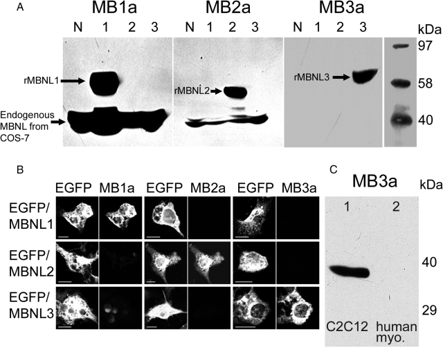
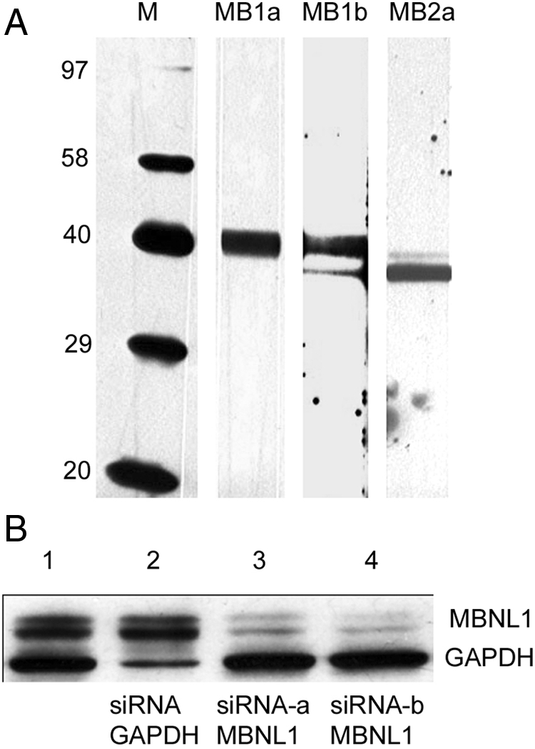
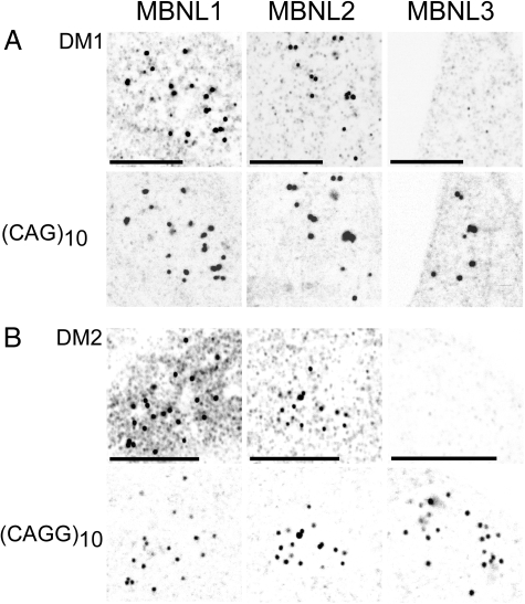
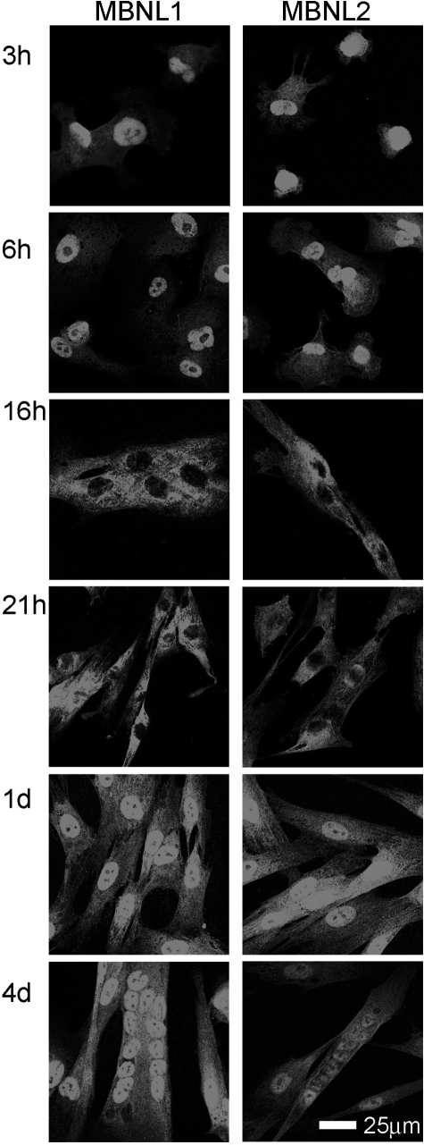
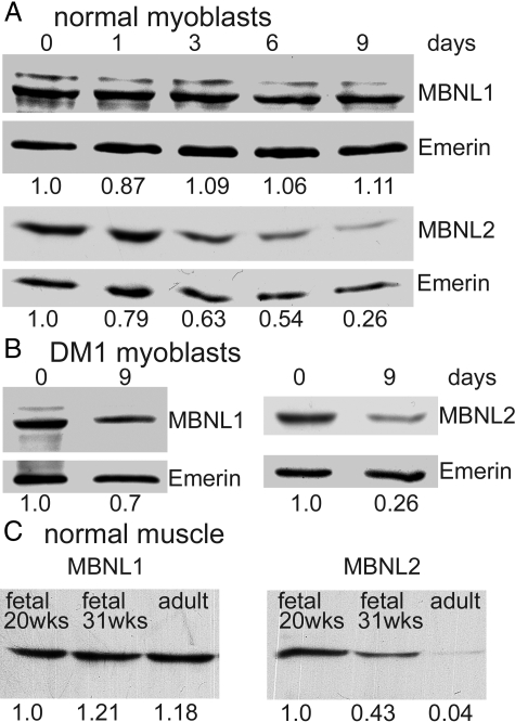
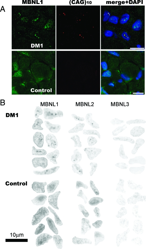
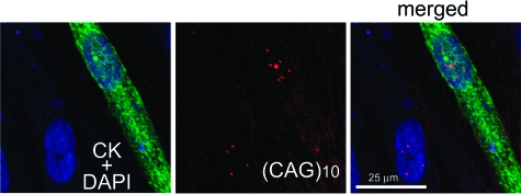
In myotonic dystrophy, muscleblind-like protein 1 (MBNL1) protein binds specifically to expanded CUG or CCUG repeats, which accumulate as discrete nuclear foci, and this is thought to prevent its function in the regulation of alternative splicing of pre-mRNAs. There is strong evidence for the role of the MBNL1 gene in disease pathology, but the roles of two related genes, MBNL2 and MBNL3, are less clear. Using new monoclonal antibodies specific for each of the three gene products, we found that MBNL2 decreased during human fetal development and myoblast culture, while MBNL1 was unchanged. In Duchenne muscular dystrophy muscle, MBNL2 was elevated in immature, regenerating fibres compared with mature fibres, supporting some developmental role for MBNL2. MBNL3 was found only in C2C12 mouse myoblasts. Both MBNL1 and MBNL2 were partially sequestered by nuclear foci of expanded repeats in adult muscle and cultured cells from myotonic dystrophy patients. In adult muscle nucleoplasm, both proteins were reduced in myotonic dystrophy type 1 compared with an age-matched control. In normal human myoblast cultures, MBNL1 and MBNL2 always co-distributed but their distribution could change rapidly from nucleoplasmic to cytoplasmic. Functional differences between MBNL1 and MBNL2 have not yet been found and may prove quite subtle. The dominance of MBNL1 in mature, striated muscle would explain why ablation of the mouse mbnl1 gene alone is sufficient to cause a myotonic dystrophy.


Figures

Figure 1
Figure 1
MBNL monoclonal antibodies are specific for each of the three isoforms in both Western blot and immunolocalization. A: COS-7 cells were transfected with EGFP constructs with MBNL1, MBNL2, or MBNL3 (1, 2, and 3) or left untransfected (N). The mAbs MB1a, MB2a, and MB3a detected their respective EGFP-fusion proteins on western blots, but did not cross-react with the other two fusion proteins. Endogenous monkey MBNL1 and MBNL2 have a lower Mr than the EGFP fusion proteins and were detected in all cell extracts. No endogenous MBNL3 was detected in COS-7 cells. B: COS-7 cells were transfected with EGFP constructs containing MBNL1, MBNL2, or MBNL3. Green fluorescence identified the transfected cells (left image of each pair). Each mAb showed intense staining of cells transfected with the corresponding MBNL isotype (right image of each pair) but did not cross-react with the other two isotypes. Some endogenous nuclear staining of COS-7 cells was seen with the MBNL1 mAb. Scale bar = 10 μm. C: Endogenous MBNL3 is detected in the mouse C2C12 myoblast cell line (lane 1), but not in human myoblast primary cultures (lane 2).
Figure 2
Figure 2
MBNL1 and MBNL2 antibodies do not cross-react with non-MBNL proteins on western blots of HeLa cells. A: MB1a mAb recognizes only MBNL1 (unresolved doublet at 40 to 42 kDa) and MB2a mAb recognizes only MBNL2. MB1b mAb recognizes both MBNL1 and MBNL2, but no non-MBNL proteins. Vertical strips of the same HeLa blot were developed with the three different antibodies for different exposure times. The intensities of the MBNL1 and MBNL2 bands stained by the same MB1b mAb may reflect the relative abundance of the two isoforms in HeLa cells. B: Two different siRNAs knocked down MBNL1 in HeLa cells without affecting the control protein glyceraldehyde-3-phosphate dehydrogenase (38.4 kDa). This shows that both bands in the doublet are authentic MBNL1. Control glyceraldehyde-3-phosphate dehydrogenase silencing did not affect MBNL1 levels.
Figure 3
Figure 3
Colocalization of MBNL1 and MBNL2 with expanded RNA repeats in nuclear foci of DM1 and DM2 cultured cells. Immunolocalization (upper image of each pair; mAbs MB1a, MB2a, and MB3a) and in situ hybridization (lower image of each pair; A: (CAG)10 probe and B: (CAGG)10 probe) in (A) DM1 myoblasts (800 CTG) and (B) DM2 skin fibroblasts (repeat size unknown). MBNL3 was not detected. Cells were fixed with acetone-methanol. Scale bar = 5 μm. Images are shown as inverted grayscale.
Figure 4
Figure 4
Changes in subcellular distribution of MBNL1 and MBNL2 during myoblast culture. Normal human myoblasts were grown on coverslips for various times before fixation with formalin, permeabilization with Triton X-100, and development with MB1a or MB2a mAbs, as described in the Methods. At day 1, the growth medium was changed to differentiation medium.
Figure 5
Figure 5
MBNL2 levels decrease during myogenesis in vitro and in vivo. A, B: Western blots with mAbs against MBNL1 (MB1a), MBNL2 (MB2a), or emerin (MANEM5) with extracts of (A) control or (B) DM1 (1300 CTG) muscle cell cultures during differentiation (0 to 9 days). To control for loading variations, the intensity of MBNL1 or MBNL2 was expressed as a ratio of intensity of the corresponding band of emerin. Day 0 was assigned a value of 1.0. With values from two control and two DM1 experiments combined (n = 4), levels of MBNL1 were not significantly different between 0 and 9 days (ratio = 0.85 ± 0.17; P = 0.2), whereas levels of MBNL2 decreased between these time points (ratio = 0.21 ± 0.10; P < 0.001; Student’s paired t-test). C: Western blots with MB1a and MB2a mAbs with extracts of control or DM1 muscle at different stages of development. Approximately equal amounts of total protein from each extract were loaded after determination of the peak areas of major protein bands on a stain gel.
Figure 6
Figure 6
Higher levels of MBNL2 in regenerating muscle fibers in a skeletal muscle biopsy from a Duchenne muscular dystrophy patient. Five regenerating fibers were identified by immunostaining with MHCn mAb (Novocastra, Newcastle, UK; 1:20) against neonatal myosin (A−C). In a serial section (D−F), nuclei in the regenerating fibers were more strongly stained for MBNL2 than nuclei in mature fibers (MB2a mAb; 1:4). The center panels show ethidium bromide staining of nuclei and the right panels are merged images. In (F), MBNL2 staining is indicated by horizontal white arrows in the nuclei of regenerating fibers and by vertical white arrows in the nuclei of mature fibers.
Figure 7
Figure 7
Fixation method affects visualization of MBNL1 in the nucleus. Cultured human DM1 myoblasts (800 CTG) were fixed and permeabilized either with acetone-methanol (A: two nuclei) or with formalin followed by Triton X-100 (B: one nucleus), as described in Materials and Methods. MBNL1 protein was detected using MB1a mAb. Scale bar = 10 μm.
Figure 8
Figure 8
Nucleoplasmic levels of both MBNL1 and MBNL2 were lower in a muscle biopsy from a congenital DM1 patient (repeat size unknown) compared with an age-matched control biopsy (with a non-DM diagnosis). Frozen sections of DM1 and control skeletal muscle biopsies, were immunostained with MB1a, MB2a, MB3a, or control mAB. A: Nuclear foci were identified by in situ hybridization in the DM1 sections but not in the control. DM1 nuclear foci of mutant RNA are also intensely stained for MBNL1. Scale bar = 10 μm. B: Nucleoplasmic staining for MBNL1 and MBNL2 outside foci is visibly reduced, but not eliminated, in DM1 sections. The ten random nuclei shown were subjected to image analysis using ImageJ software (Table 2).
Figure 9
Figure 9
Differentiation of DM1 myoblasts (800 CTG) leads to a doubling in the number of foci per nucleus. Creatine kinase (CK) antibody was used to distinguish a CK-positive differentiated cell and a CK-negative myoblast in the same field (left, green). In situ hybridization was used to identify nuclear foci of mutant RNA in the same cells (center, red). Nuclei are counterstained with DAPI (blue). The CK-negative myoblast contains 4 foci and the CK-positive myoblast nucleus contains 7 to 10 foci.

Similar articles

Cited by

References

    1. Harper PS. London: WB Saunders; Myotonic Dystrophy. (ed 3) 2004
    1. Ranum LP, Day JW. Myotonic dystrophy: rNA pathogenesis comes into focus. Am J Hum Genet. 2004;7:793–804. - PMC - PubMed
    1. Day JW, Ranum LP. RNA pathogenesis of the myotonic dystrophies. Neuromuscul Disord. 2005;15:5–16. - PubMed
    1. Machuca-Tzili L, Brook D, Hilton-Jones D. Clinical and molecular aspects of the myotonic dystrophies: a review. Muscle Nerve. 2005;32:1–18. - PubMed
    1. Brook JD, McCurrach ME, Harley HG, Buckler AJ, Church D, Aburatani H, Hunter K, Stanton VP, Thirion JP, Hudson T, Sohn R, Zemelman B, Snell RG, Rundle SA, Crow S, Davies J, Shelbourne P, Buxton J, Jones C, Juvonen V, Johnson K, Harper PS, Shaw DJ, Housman DE. Molecular basis of myotonic dystrophy: expansion of a trinucleotide (CTG) repeat at the 3′ end of a transcript encoding a protein kinase family member. Cell. 1992;68:799–808. - PubMed

Publication types

MeSH terms