. 2009 Jan 30;104(2):210-8, 21p following 218.
doi: 10.1161/CIRCRESAHA.108.181040. Epub 2008 Dec 18.

Macrophages generate reactive oxygen species in response to minimally oxidized low-density lipoprotein: toll-like receptor 4- and spleen tyrosine kinase-dependent activation of NADPH oxidase 2

Affiliations

Macrophages generate reactive oxygen species in response to minimally oxidized low-density lipoprotein: toll-like receptor 4- and spleen tyrosine kinase-dependent activation of NADPH oxidase 2

Yun Soo Bae et al. Circ Res. .

Abstract

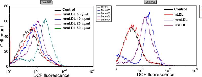
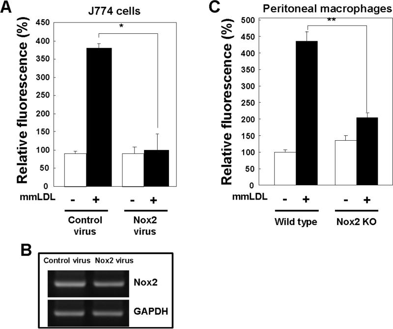
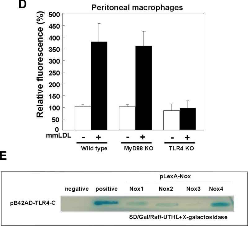
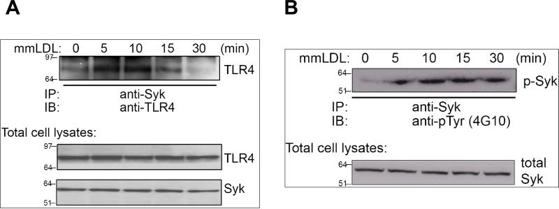
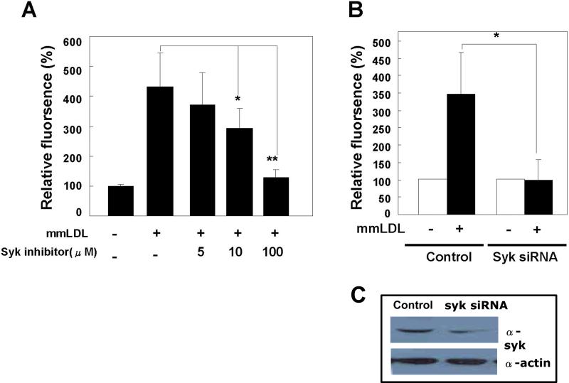
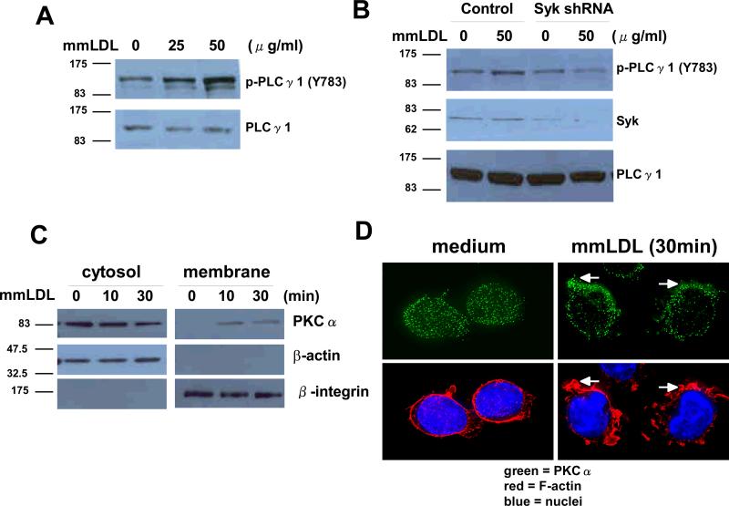
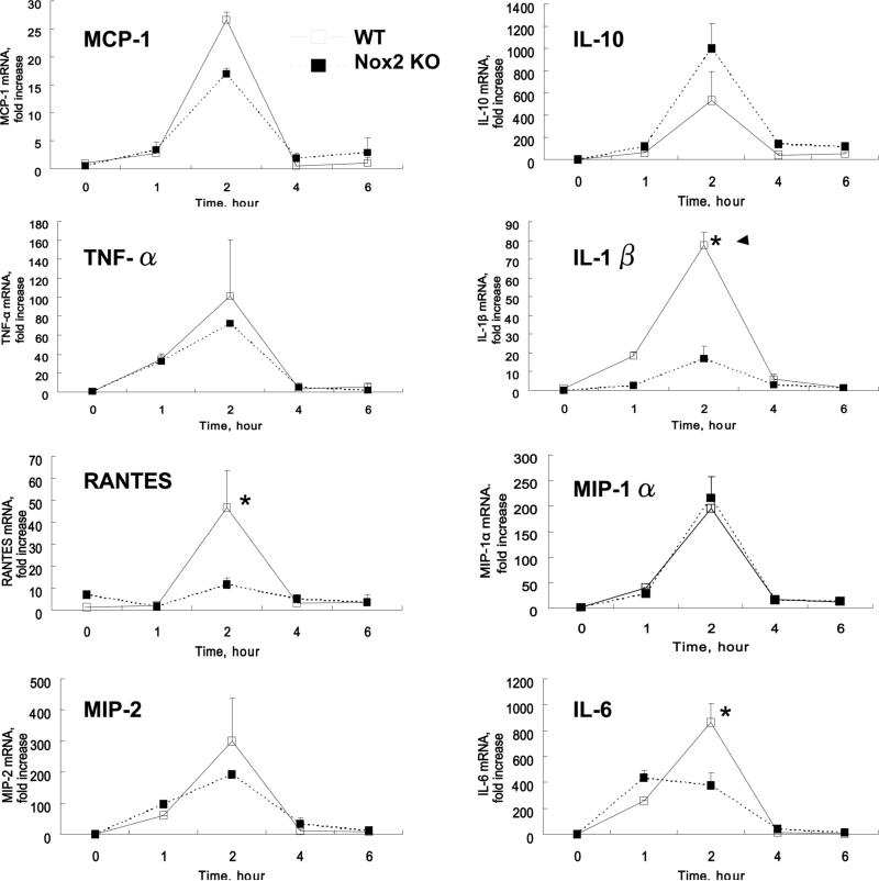
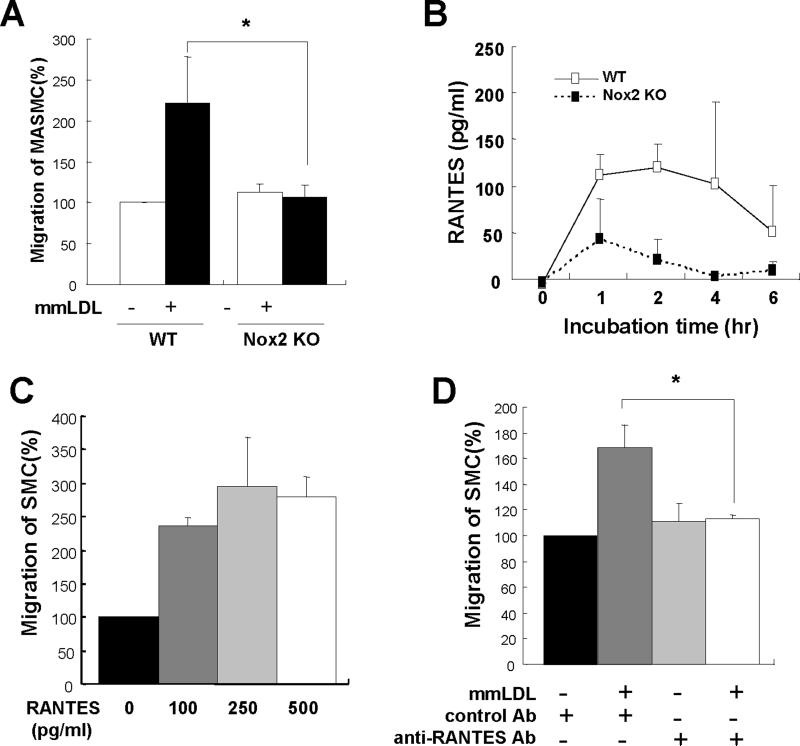
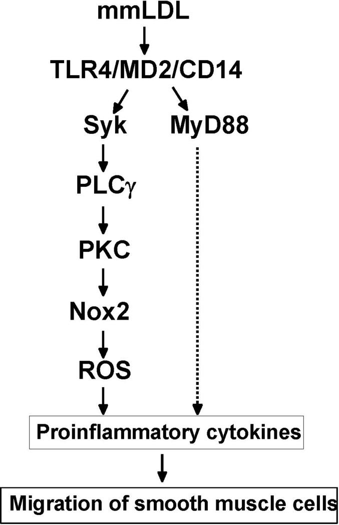
Oxidative modification of low-density lipoprotein (LDL) plays a causative role in the development of atherosclerosis. In this study, we demonstrate that minimally oxidized LDL (mmLDL) stimulates intracellular reactive oxygen species (ROS) generation in macrophages through NADPH oxidase 2 (gp91phox/Nox2), which, in turn, induces production of RANTES and migration of smooth muscle cells. Peritoneal macrophages from gp91phox/Nox2(-/-) mice or J774 macrophages in which Nox2 was knocked down by small interfering RNA failed to generate ROS in response to mmLDL. Because mmLDL-induced cytoskeletal changes were dependent on Toll-like receptor (TLR)4, we analyzed ROS generation in peritoneal macrophages from wild-type, TLR4(-/-), or MyD88(-/-) mice and found that mmLDL-mediated ROS was generated in a TLR4-dependent, but MyD88-independent, manner. Furthermore, we found that ROS generation required the recruitment and activation of spleen tyrosine kinase (Syk) and that mmLDL also induced phospholipase PLCgamma1 phosphorylation and protein kinase C membrane translocation. Importantly, the phospholipase Cgamma1 phosphorylation was reduced in J774 cells expressing Syk-specific short hairpin RNA. Nox2 modulated mmLDL activation of macrophages by regulating the expression of proinflammatory cytokines interleukin-1beta, interleukin-6, and RANTES. We showed that purified RANTES was able to stimulate migration of mouse aortic smooth muscle cells and addition of neutralizing antibody against RANTES abolished the migration of mouse aortic smooth muscle cells stimulated by mmLDL-stimulated macrophages. These results suggest that mmLDL induces generation of ROS through sequential activation of TLR4, Syk, phospholipase Cgamma1, protein kinase C, and gp91phox/Nox2 and thereby stimulates expression of proinflammatory cytokines. These data help explain mechanisms by which endogenous ligands, such as mmLDL, can induce TLR4-dependent, proatherogenic activation of macrophages.


Figures

Figure 1
Figure 1
MmLDL stimulates ROS generation in J774 macrophages. (A) J774 cells were incubated for 10 min with indicated concentrations of mmLDL, followed by a 10 min incubation with DCF-DA. The generation of ROS (H2O2) was then monitored by FACS analysis as an increase in DCF fluorescence. Fluorescence was analyzed in 10,000 cells with excitation at 488nm and emission at 530nm. (B) J774 cells were incubated with equal amounts (50μg/ml) of native LDL (nLDL), minimally oxidized LDL (mmLDL), or extensively oxidized LDL (OxLDL) for 10 min and ROS generation was measured as in A.
Figure 2
Figure 2
Effect of Nox2 depletion on mmLDL–induced ROS generation. (A) J774 cells were infected with either a retrovirus encoding Nox2-specific siRNA or a retrovirus encoding scrambled siRNA. After 48 hours, cells were stimulated with mmLDL for 10 min, and the generation of H2O2 was monitored by FACS as in Fig. 1. Data are means ± S.E. of mean fluorescence intensity from three independent experiments. *Comparison to the value of control group, p<0.005. (B) Nox2 mRNA expression was assessed in total RNA by RT-PCR as described in Methods. GAPDH served as the loading control. (C and D) Resident peritoneal macrophages were obtained from C57BL/6 wild type, Nox2−/−, MyD88−/− and TLR4−/− mice, stained with a PE-labeled CD11b antibody to select for macrophages and then incubated with mmLDL (50μg/ml) for 10 min, followed by a 10 min incubation with DCF-DA. DCF fluorescence was then measured by FACS in the population of CD11b-positive cells. Data are means ± S.E. of mean fluorescence intensity from three independent experiments. **Comparison to the value of control group, p<0.0001 and ***p<0.01 wild type vs. TLR4−/− macrophages (E) Interactions of the COOH-terminal region of TLR4 with the COOH-terminal domains of Nox1, Nox2, Nox3, or Nox4 were estimated using a yeast two-hybrid assay (see Methods). The intensity of blue color indicates the expression levels of LacZ and corresponds to the affinity of TLR4 binding with Nox isozymes.
Figure 2
Figure 2
Effect of Nox2 depletion on mmLDL–induced ROS generation. (A) J774 cells were infected with either a retrovirus encoding Nox2-specific siRNA or a retrovirus encoding scrambled siRNA. After 48 hours, cells were stimulated with mmLDL for 10 min, and the generation of H2O2 was monitored by FACS as in Fig. 1. Data are means ± S.E. of mean fluorescence intensity from three independent experiments. *Comparison to the value of control group, p<0.005. (B) Nox2 mRNA expression was assessed in total RNA by RT-PCR as described in Methods. GAPDH served as the loading control. (C and D) Resident peritoneal macrophages were obtained from C57BL/6 wild type, Nox2−/−, MyD88−/− and TLR4−/− mice, stained with a PE-labeled CD11b antibody to select for macrophages and then incubated with mmLDL (50μg/ml) for 10 min, followed by a 10 min incubation with DCF-DA. DCF fluorescence was then measured by FACS in the population of CD11b-positive cells. Data are means ± S.E. of mean fluorescence intensity from three independent experiments. **Comparison to the value of control group, p<0.0001 and ***p<0.01 wild type vs. TLR4−/− macrophages (E) Interactions of the COOH-terminal region of TLR4 with the COOH-terminal domains of Nox1, Nox2, Nox3, or Nox4 were estimated using a yeast two-hybrid assay (see Methods). The intensity of blue color indicates the expression levels of LacZ and corresponds to the affinity of TLR4 binding with Nox isozymes.
Figure 3
Figure 3
Syk association with TLR4 and phosphorylation of Syk. J774 cells were incubated with mmLDL for the indicated times and the incubations were terminated by the addition of a lysis buffer. Cell lysates were subjected to immunoprecipitation with an antibody against Syk and then the immune complexes were directly subjected to immunoblot analysis with an antibody against TLR4 (A) or phosphotyrosine (4G10) (B). Total cell lysates were used for the control of Syk and TLR4 load. IP and IB designate immunoprecipitation and immunoblot, respectively.
Figure 4
Figure 4
Role of Syk in mmLDL-induced ROS generation. (A) J774 cells were pretreated for 30 minutes with indicated concentrations of the Syk inhibitor, piceatannol, and then incubated with 50 μg/ml mmLDL for additional 10 min. ROS was measured as in Fig. 1. Data are means ± S.E. of mean fluorescence intensities from three independent experiments. *, p<0.02 and **, p<0.001 vs. control. (B) J774 cells were transfected with either Syk-specific shRNA or control shRNA. After 48 hours, the cells were stimulated with mmLDL (50 μg/ml) for 10 min and ROS were measured. Data are means ± S.E. of mean fluorescence intensities from five independent experiments. *Comparison to the value of control group, P<0.01. (C) The Syk knockdown was confirmed in cell lysates subjected to western blot analysis with antibodies against Syk and β-actin (loading control).
Figure 5
Figure 5
MmLDL-induced activation of PLCγ1 and membrane translocation of PKCα. (A) J774 cells were stimulated with mmLDL (25 or 50 μg/ml) for 10 min. Cell lysates from each sample were then prepared and subjected to western blot analysis with antibodies to PLCγ1 or phospho-specific PLCγ1 (Y783). (B) J774 cells were transfected with either Syk-specific shRNA or control shRNA. After 48 hours, the cells were stimulated with mmLDL (50 μg/ml) for 10 min. Cell lysates from each sample were then subjected to western blot analysis with antibodies against Syk, PLCγ1 or phospho-specific PLCγ1 (Y783). (C) J774 cells were stimulated with mmLDL (50 μg/ml) for indicated times and subjected to cellular fractionation (see Methods). Density fractions were immunoblotted with antibodies against PKCα, β-actin (cytosolic marker) and β2-integrin (membrane marker). (D) J774 cells were incubated with medium alone or medium plus mmLDL (50 μg/ml) for 30min. PKCα localization was visualized with a primary anti-PKCα antibody and a secondary FITC-labeled antibody. Cells were also stained with TRITC-phalloidin and Hoechst 33258 to visualize F-actin and nuclei, respectively.
Figure 6
Figure 6
MmLDL-induced expression of cytokines in peritoneal resident macrophages. Peritoneal resident macrophages were isolated from wild type and Nox2−/− mice and stimulated with mmLDL (50 μg/ml) or media only for indicated times. Total RNA was isolated, reverse transcribed, and quantified by real-time PCR with respective primers specific for each cytokine and GAPDH. The data are presented as a fold increase in mRNA levels in mmLDL-stimulated cells over the levels in non-stimulated cells. *, p<0.05 wild type vs. Nox2−/− macrophages.
Figure 7
Figure 7
RANTES-dependent MASMC migration. (A) Mouse aortic smooth muscle cells (MASMC) (8 × 10³ cells in 100 μl media) were added to the upper chamber and peritoneal macrophages from wild type or Nox2−/− mice in the lower chamber. First, macrophages were activated with media alone or 50 μg/ml mmLDL for 2h, and then upper and lower chambers were assembled. After 18h incubation, the membrane in the upper chamber was recovered, fixed, stained and the number of migrated cells was counted. Data are from five independent experiments. *, p<0.05 mmLDL/WT vs. mmLDL/Nox2−/−. (B) Analysis of RANTES protein secretion into cell culture media following macrophage activation with mmLDL (50 μg/ml) for 0, 1, 2, 4 or 6 hours. (C) Mouse aortic smooth muscle cells (MASMC) (8 × 10³ cells in 100 μl media) and RANTES (0, 100, 250 or 500 pg/ml) were added to the upper chamber and lower chamber, respectively, and incubated for 18h, and the numbers of migrated cells were counted, (D) Blocking experiments were performed by incubating the mmLDL-stimulated macrophages (wild type) with 100 ng/ml of the neutralizing antibody against RANTES (R&D systems) or with the isotype-matched control antibody in the lower chamber. The number of migrated cells was determined as above. Data are from three independent experiments. *, p<0.05 vs. mmLDL/anti-RANTES antibody.
Figure 8
Figure 8
A proposed signaling pathway of mmLDL-induced, TLR4/Syk-dependent ROS generation and proinflammatory cytokine expression.

Similar articles

Cited by

References

    1. Rhee SG. Cell signaling. H2O2, a necessary evil for cell signaling. Science. 2006;312:1882–1883. - PubMed
    1. Bedard K, Krause KH. The NOX family of ROS-generating NADPH oxidases: physiology and pathophysiology. Physiol Rev. 2007;87:245–313. - PubMed
    1. Zhang X, Shan P, Jiang G, Cohn L, Lee PJ. Toll-like receptor 4 deficiency causes pulmonary emphysema. J Clin Invest. 2006;116:3050–3059. - PMC - PubMed
    1. Park HS, Jung HY, Park EY, Kim J, Lee WJ, Bae YS. Cutting edge: direct interaction of TLR4 with NAD(P)H oxidase 4 isozyme is essential for lipopolysaccharide-induced production of reactive oxygen species and activation of NF-kappa B. J Immunol. 2004;173:3589–3593. - PubMed
    1. Kawai T, Akira S. TLR signaling. Seminars in Immunol. 2007;19:24–32. - PubMed

Publication types

MeSH terms

Substances