. 2009 Feb 13;104(3):380-7.
doi: 10.1161/CIRCRESAHA.108.180695. Epub 2008 Dec 18.

Heparanase alters arterial structure, mechanics, and repair following endovascular stenting in mice

Affiliations

Heparanase alters arterial structure, mechanics, and repair following endovascular stenting in mice

Aaron B Baker et al. Circ Res. .

Abstract

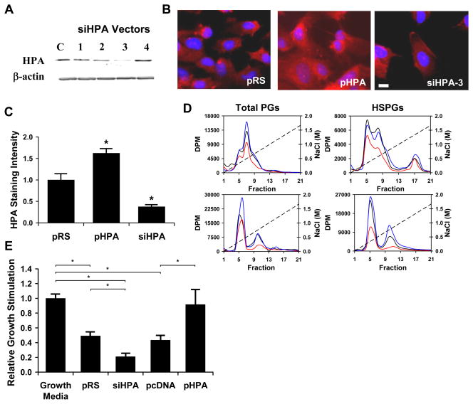
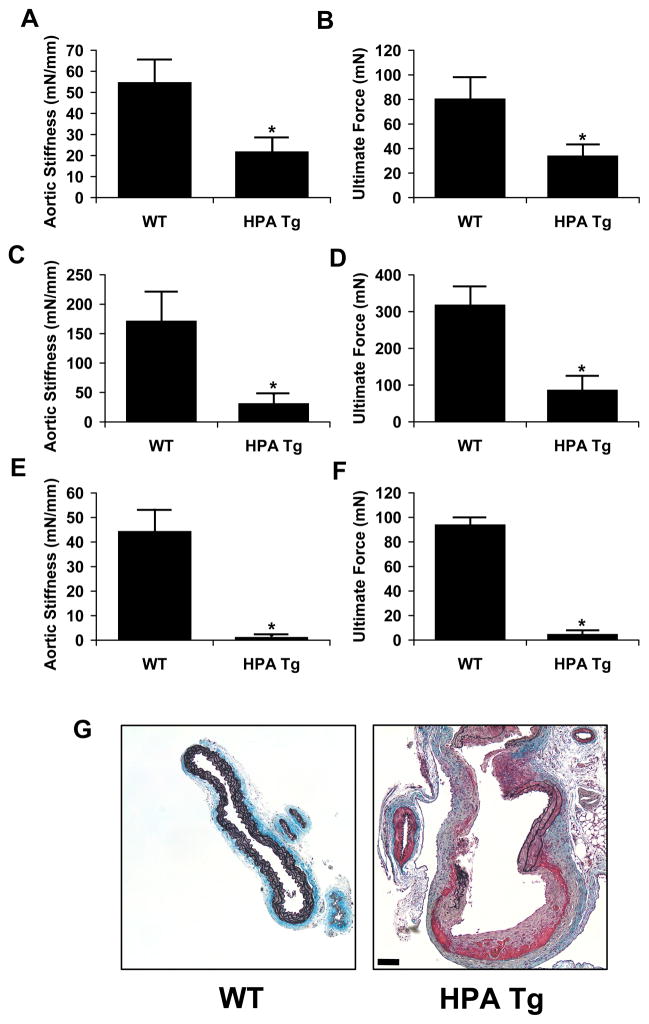
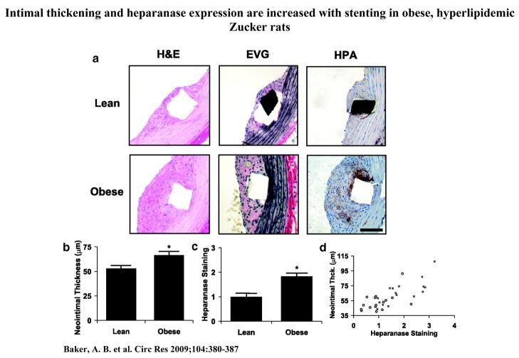
Heparan sulfate proteoglycans (HSPGs) are potent regulators of vascular remodeling and repair. Heparanase is the major enzyme capable of degrading heparan sulfate in mammalian cells. Here we examined the role of heparanase in controlling arterial structure, mechanics, and remodeling. In vitro studies supported that heparanase expression in endothelial cells serves as a negative regulator of endothelial inhibition of vascular smooth muscle cell (vSMC) proliferation. Arterial structure and remodeling to injury were also modified by heparanase expression. Transgenic mice overexpressing heparanase had increased arterial thickness, cellular density, and mechanical compliance. Endovascular stenting studies in Zucker rats demonstrated increased heparanase expression in the neointima of obese, hyperlipidemic rats in comparison to lean rats. The extent of heparanase expression within the neointima strongly correlated with the neointimal thickness following injury. To test the effects of heparanase overexpression on arterial repair, we developed a novel murine model of stent injury using small diameter self-expanding stents. Using this model, we found that increased neointimal formation and macrophage recruitment occurs in transgenic mice overexpressing heparanase. Taken together, these results support a role for heparanase in the regulation of arterial structure, mechanics, and repair.


Figures

Figure 1
Figure 1
Heparanase knock down and overexpression modulates endothelial inhibition of vascular smooth muscle cell (vSMC) proliferation. (a) Expression of heparanase identified by Western blotting in cells transfected with a vector expressing siRNA specific for mammalian heparanase (siHPA). Four siHPA were evaluated and the one that most effectively reduced heparanase was used in all subsequent experiments. A scrambled sequence (pRS) served as control. (b) Immunocytochemically identified heparanase (Bar = 10 μm) in cells transfected with pHPA, siHPA or pRS. (c) Quantification of cellular staining for heparanase in transfected cells. Values are based on measurement of intensity of the average of ten cells from five independent experiments (n=5). *Statistically significant difference from control group (p < 0.05). (d) Measurement of heparanase’s effect on total glycosaminoglycans and heparan sulfate in endothelial cells. Endothelial cell HSPGs were metabolically labeled using 3H-glucosamine. Cell lysates and conditioned media were then analyzed for glycosaminoglycan content using ion exchange chromatography. Heparanase overexpression caused a reduction in cellular and soluble HSPGs in endothelial cells. Conversely, reduction of heparanase expression in endothelial cells by siRNA led to an increase in both cellular and soluble HSPGs. Plots are the average of three independent analyses. Black line = pRS; red line = pHPA; blue line = siHPA. (e) Heparanase regulates endothelial inhibition of vSMC proliferation. Conditioned media from endothelial cells that were transfected with pcDNA, pHPA, siHPA or pRS vectors was harvested and applied to cultures of vSMCs. Bars represent the relative growth of vSMC in the presence of conditioned media of endothelial cells transfected with the indicated vectors as measured by 3H-thymidine incorporation. *Statistically significant difference between groups (p < 0.05).
Figure 2
Figure 2
Transgenic mice overexpressing heparanase demonstrate altered arterial structure and cellular density. Thoracic aortae were harvested from transgenic and wild-type animals (n = 8 for each group), formalin fixed, paraffin embedded and sectioned. Aortic sections were taken from four animals of each group at three different locations on the aorta. (a) Histological analysis of heparanase transgenic (HPA Tg) and wild-type (WT) mice stained with H&E and elastic Movat pentachrome stain (EMP). (b) Aortic thickness is increased in heparanase transgenic mice. *Statistically significant difference between samples (n = 4, p < 0.05). Bar = 50 μm.
Figure 3
Figure 3
Overexpression of heparanase alters the mechanical compliance and ultimate strength of the mouse aorta. The thoracic aortae were harvested from transgenic and wild-type animals and mechanical properties measured using a Bose BioDynamic test instrument. (a) Aortic stiffness expressed as the slope of the force-displacement curve was reduced in HPA Tg animals. (b) Average aortic ultimate strength was reduced in mice expressing the heparanase transgene. (c) Circumferential aortic stiffness of wild type and HPA Tg animals. (d) Circumferential ultimate force for aortae isolated from wild type and HPA Tg animals. (e) Circumferential stiffness of the elastin network remaining after digestion with NaOH solution. (f) Circumferential ultimate strength of the elastin network remaining after digestion with NaOH solution. (g) Example of a local, spontaneous aneurysm found in the aorta of a transgenic heparanase mouse compared to a wild type control. The sample was stained with elastic Movat’s pentachrome stain. Bar = 50 μm.
Figure 4
Figure 4
Intimal thickening and heparanase expression are increased with stenting in obese, hyperlipidemic Zucker rats. Endovascular stenting was performed through minimally invasive femoral access. (a) Histochemical staining and immunohistochemical staining of lean and obese Zucker rat aorta, 14 days after stent placement (n=16). EVG = Elastic Verhoff Van Geisen staining. Bar = 50 μm. (b) Analysis of neointimal thickness in lean and obese Zucker rats. (c) Quantitative analysis of heparanase in the neointima of lean and obese Zucker rats. (d) Heparanase expression and neointimal proliferation in response to endovascular stenting are enhanced in obese Zucker rats (■) compared to lean controls (○).The expression of heparanase within the neointima correlated strongly with the final neointimal area, giving a correlation of R=0.50 (p < 0.001) for lean rats and R=0.77 (p < 0.001) for fatty rats. *Statistically significant difference between samples (p < 0.05).
Figure 5
Figure 5
Abdominal stenting of heparanase transgenic and wild type mice was performed using femoral access with small diameter self-expanding stents. Stents were harvested after 14 days and processed for histological analysis in resin sections. (a) Lateral x-ray of stent placement in the abdominal aorta of the mouse. The stent is visible just below the spine (marked with arrow). (b) Neointimal formation in response to vascular injury with endovascular staining is increased in heparanase transgenic mice. Staining for Mac-1 was increased in heparanase transgenic mice. Bar = 50 μm. (c–e) Morphological analysis of the stented arteries showed increased intimal area, intima to medial ratio and no change in media area for heparanase transgenic mice. (f) Concentration of MCP-1 in arterial lysates as measured by ELISA assay. The arteries were harvested one hour following arterial injury (n=4). *Statistically significant difference between all samples (p < 0.05).
Figure 5
Figure 5
Abdominal stenting of heparanase transgenic and wild type mice was performed using femoral access with small diameter self-expanding stents. Stents were harvested after 14 days and processed for histological analysis in resin sections. (a) Lateral x-ray of stent placement in the abdominal aorta of the mouse. The stent is visible just below the spine (marked with arrow). (b) Neointimal formation in response to vascular injury with endovascular staining is increased in heparanase transgenic mice. Staining for Mac-1 was increased in heparanase transgenic mice. Bar = 50 μm. (c–e) Morphological analysis of the stented arteries showed increased intimal area, intima to medial ratio and no change in media area for heparanase transgenic mice. (f) Concentration of MCP-1 in arterial lysates as measured by ELISA assay. The arteries were harvested one hour following arterial injury (n=4). *Statistically significant difference between all samples (p < 0.05).

Comment in

References

    1. Cohen JD. Overview of physiology, vascular biology, and mechanisms of hypertension. J Manag Care Pharm. 2007;13:S6–8. - PMC - PubMed
    1. Libby P. Atherosclerosis: disease biology affecting the coronary vasculature. Am J Cardiol. 2006;98:3Q–9Q. - PubMed
    1. Weintraub WS. The pathophysiology and burden of restenosis. Am J Cardiol. 2007;100:3K–9K. - PubMed
    1. Karnovsky MJ, Edelman ER. Heparin/heparan sulphate regulation of vascular smooth muscle behaviour. In: Page CP, Black JL, editors. Airways and Vascular Remodeling in Asthma and Cardiovascular Disease: Implications for Therapeutic Intervention. New York: Academic Press; 1994. pp. 45–70.
    1. Michiels C. Endothelial cell functions. J Cell Physiol. 2003;196:430–443. - PubMed

Publication types

MeSH terms