. 2009 Mar;27(3):753-60.
doi: 10.1634/stemcells.2008-0831.

Tumor immunotherapy using gene-modified human mesenchymal stem cells loaded into synthetic extracellular matrix scaffolds

Affiliations
Free PMC article

Tumor immunotherapy using gene-modified human mesenchymal stem cells loaded into synthetic extracellular matrix scaffolds

Marta Compte et al. Stem Cells. 2009 Mar.
Free PMC article

Abstract

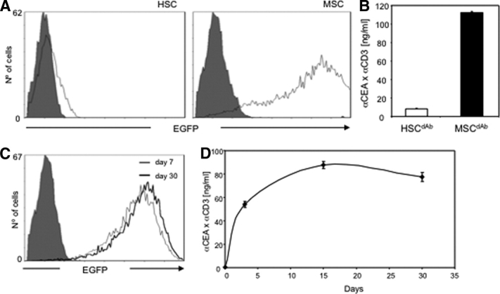
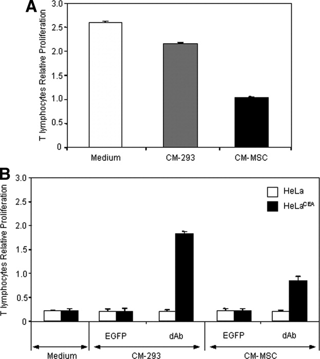
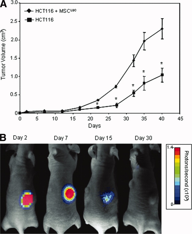
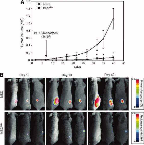
Mesenchymal stem cells (MSCs) are appealing as gene therapy cell vehicles given their ease of expansion and transduction. However, MSCs exhibit immunomodulatory and proangiogenic properties that may pose a risk in their use in anticancer therapy. For this reason, we looked for a strategy to confine MSCs to a determined location, compatible with a clinical application. Human MSCs genetically modified to express luciferase (MSC(luc)), seeded in a synthetic extracellular matrix (sECM) scaffold (sentinel scaffold) and injected subcutaneously in immunodeficient mice, persisted for more than 40 days, as assessed by bioluminescence imaging in vivo. MSCs modified to express a bispecific alpha-carcinoembryonic antigen (alphaCEA)/alphaCD3 diabody (MSC(dAb)) and seeded in an sECM scaffold (therapeutic scaffolds) supported the release of functional diabody into the bloodstream at detectable levels for at least 6 weeks after implantation. Furthermore, when therapeutic scaffolds were implanted into CEA-positive human colon cancer xenograft-bearing mice and human T lymphocytes were subsequently transferred, circulating alphaCEA/alphaCD3 diabody activated T cells and promoted tumor cell lysis. Reduction of tumor growth in MSC(dAb)-treated mice was statistically significant compared with animals that only received MSC(luc). In summary, we report here for the first time that human MSCs genetically engineered to secrete a bispecific diabody, seeded in an sECM scaffold and implanted in a location distant from the primary tumor, induce an effective antitumor response and tumor regression.


Figures

Figure 1
Figure 1
Transduction of HSCs and mesenchymal stem cells (MSCs) with dAb.EGFP-encoding lentivirus (LentidAb) at a multiplicity of infection of 10. (A, B): Analysis of EGFP expression (A) and secretion of α-CEA/αCD3 diabody into the cell culture supernatant (B) 72 hours after transduction. (C, D): Transgene expression stability in MSCs (MSCdAb): expression of EGFP (C) and secretion of α-CEA/αCD3 dAb (D). Abbreviations: α-CEA, α-carcinoembryonic antigen; dAb, diabody; EGFP, enhanced green fluorescent protein; HSC, hematopoietic stem cells; MSC, mesenchymal stem cell; MSCdAb, mesenchymal stem cells after infection with LentidAb.
Figure 2
Figure 2
Inhibitory effect of MSCs on T lymphocyte proliferation. (A): CM-MSC inhibits the proliferation of human T lymphocytes following polyclonal stimuli. (B): CM-MSCdAb inhibits the specific proliferation of T lymphocytes induced by αCEA/αCD3 diabody in presence of CEA-positive cells. Approximately 105 human T lymphocytes were stimulated (effector/target ratio = 5:1) with irradiated CEA-negative (HeLa) or CEA-positive (HeLaCEA) target cells in the presence of medium or cell-free conditioned medium from cultures of HEK-293 or MSCs transduced with either LentiEGFP or LentidAb. As controls, effector and target cells were cultured alone (data not shown). Proliferation was analyzed after 72 hours of culture. Abbreviations: CEA, carcinoembryonic antigen; CM-293, conditioned medium from HEK-293 cells; CM-MSC, conditioned medium from mesenchymal stem cells; CM-MSCdAb, CM from mesenchymal stem cells transduced with LentidAb; dAb, diabody; EGFP, enhanced green fluorescent protein.
Figure 3
Figure 3
Effect of intratumoral MSCs on human colorectal carcinoma xenograft growth. (A): Tumor volume measurements of HCT-116 cells (2 × 10⁶) injected s.c. into nude mice (n = 4 mice per group) with or without 5 × 10⁵ MSCLuc. (B): The persistence of viable functional MSCLuc in the tumors was assessed by bioluminescence imaging (BLI). BLI of a representative mouse is shown 2, 7, 15, and 30 days after implantation of HCT-116 and MSCLuc cells. Abbreviation: MSCLUC, mesenchymal stem cells infected with luciferase-encoding lentivirus.
Figure 4
Figure 4
Establishment and characterization of MSCs-seeded Extracel-X scaffolds. (A): The persistence of viable functional MSCLuc in the scaffolds was assessed by bioluminescence imaging (BLI) 1, 7, 30, and 42 days after implantation. Shown is BLI of a representative mouse (total, n = 3 mice per group). (B): Plasma concentration of αCEA/αCD3 diabody in mice implanted with MSCdAb-embedded scaffolds. Abbreviation: αCEA, α-carcinoembryonic antigen.
Figure 5
Figure 5
Human colorectal carcinoma xenograft growth in nude mice bearing MSCdAb-seeded scaffold. (A): Tumor volume measurements of HCT-116 cells (2 × 10⁶) infected with luciferase-encoding lentivirus (HCT-116Luc) injected s.c. into the dorsal skin of nude mice (n = 3 mice per group). Five days after tumor implantation, MSCs or MSCdAb (1 × 10⁶) were seeded in a synthetic extracellular matrix Extracel-X scaffold and inoculated s.c. in the ventral area. Two days later, animals received an i.v. injection of 2 × 10⁶ human unfractionated peripheral blood lymphocytes. (B): Bioluminescence imaging assay for monitoring tumor growth in vivo, at days 7, 30, and 42 after tumor implantation. Abbreviations: dAb, diabody; MSC, mesenchymal stem cell.

References

    1. Pittenger MF. Mesenchymal stem cells from adult bone marrow. Methods Mol Biol. 2008;449:27–44. - PubMed
    1. Kumar S, Chanda D, Ponnazhagan S. Therapeutic potential of genetically modified mesenchymal stem cells. Gene Ther. 2008;15:711–715. - PubMed
    1. Aboody KS, Najbauer J, Danks MK. Stem and progenitor cell-mediated tumor selective gene therapy. Gene Ther. 2008;15:739–752. - PubMed
    1. Ren C, Kumar S, Chanda D, et al. Therapeutic potential of mesenchymal stem cells producing interferon-α in a mouse melanoma lung metastasis model. Stem Cells. 2008;26:2332–2338. - PMC - PubMed
    1. Nakamizo A, Marini F, Amano T, et al. Human bone marrow-derived mesenchymal stem cells in the treatment of gliomas. Cancer Res. 2005;65:3307–3318. - PubMed

Publication types

MeSH terms