. 2009 Jan 21;28(2):112-22.
doi: 10.1038/emboj.2008.268. Epub 2008 Dec 18.

Functional specialization of transcription elongation factors

Affiliations

Functional specialization of transcription elongation factors

Georgiy A Belogurov et al. EMBO J. .

Abstract

Elongation factors NusG and RfaH evolved from a common ancestor and utilize the same binding site on RNA polymerase (RNAP) to modulate transcription. However, although NusG associates with RNAP transcribing most Escherichia coli genes, RfaH regulates just a few operons containing ops, a DNA sequence that mediates RfaH recruitment. Here, we describe the mechanism by which this specificity is maintained. We observe that RfaH action is indeed restricted to those several operons that are devoid of NusG in vivo. We also show that RfaH and NusG compete for their effects on transcript elongation and termination in vitro. Our data argue that RfaH recognizes its DNA target even in the presence of NusG. Once recruited, RfaH remains stably associated with RNAP, thereby precluding NusG binding. We envision a pathway by which a specialized regulator has evolved in the background of its ubiquitous paralogue. We propose that RfaH and NusG may have opposite regulatory functions: although NusG appears to function in concert with Rho, RfaH inhibits Rho action and activates the expression of poorly translated, frequently foreign genes.


Figures
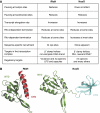
Figure 1
Figure 1
RfaH and NusG: similarities and differences. (A) Comparison of the functional properties. RfaH and NusG likely bind to the same site on RNAP (Belogurov et al, 2007 and unpublished data), the β′ CH and increase the transcript elongation rate. In contrast to RfaH, NusG increases Rho-dependent termination, presumably by stabilizing a quarternary Rho–TEC complex (Nehrke and Platt, 1994). In addition, NusG does not affect RNAP paused at the hairpin-dependent pause sites, whereas RfaH facilitates transcription similarly through both the hairpin-dependent and -independent signals (Artsimovitch and Landick, 2000, 2002). NusG also participates in the formation of multi-component transcription antitermination complexes (Mason et al, 1992; Squires et al, 1993) where it may make specific contacts to several transcription factors. Finally, NusG does not exhibit any DNA sequence specificity and does not bind to the ops-paused TECs in vitro. (B) Structures of the E. coli RfaH (Belogurov et al, 2007) and a model of the E. coli NusG (Steiner et al, 2002).
Figure 2
Figure 2
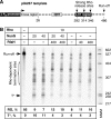
RfaH eliminates the NusG effect on Rho-dependent termination. (A) Transcript generated on a linear pIA267 DNA template; transcription start site (+1), ops, Rho-dependent RNA release sites, and transcript end are indicated. (B) Halted [α32P]GMP TECs were formed at 40 nM with E. coli RNAP. Rho, RfaH, and NusG were added where indicated. Sizes of the [γ32P]ATP pBR322 MspI fragments used as molecular markers are indicated on the right. Positions of the run-off (RO) transcript and the NusG-enhanced termination site (T*) are shown by arrows; position of the Rho-dependent termination region is indicated by a bracket.
Figure 3
Figure 3
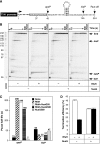
Interplay between RfaH and NusG during elongation in vitro. (A) Transcript generated from the T7A1 promoter on a linear pIA349 DNA template; start site (+1), ops and his pause sites, and RO are indicated. (B) Single-round pause assay. Halted [α32P]CMP G37 TECs formed with E. coli RNAP were incubated with RfaH (50 nM) and/or NusG (500 nM), as indicated and transcription was restarted (see Materials and methods). Positions of the G37, ops and his paused RNAs and RO transcripts are indicated with arrows. Sizes of the pBR322 MspI fragments are indicated on the left. (C) Quantification of the data shown in (B) and assays performed with 50 nM each RfaH and NusG; assays were repeated three or more times for each protein combination. (D) Termination assay was performed as described in Carter et al (2004); the error bars are the standard deviations from four independent measurements.
Figure 4
Figure 4
The RfaH NTD resists NusG competition. (A, B) The pause assay on template shown in Figure 3A. NusG was present at 500 nM, RfaHNTD was present at 50 nM. (B) Quantification of the opsP (triangles) and hisP (circles) pause RNAs from data shown in (A). Open symbols, NTD alone; filled symbols, NTD and NusG together. Pause half-lives are indicated below the gel panel. (C) Two possible fates of the RfaH-modified TEC. In the absence of translation in vivo (or in vitro), RfaH dissociates from the complex and re-establishes the interdomain interface. With concurrent translation, the CTD is sequestered, allowing the NTD to stay bound.
Figure 5
Figure 5
RfaH and NusG are targeted to different operons. (A) Gene-averaged RfaH ChIP-on-chip signal as a ratio to RNAP signal plotted as a function of log2 RNAP ChIP-on-chip signal (Materials and methods). Small black circles, protein-coding genes; red circles, rRNA genes; green circles, tRNA genes; orange circles, small RNA genes. The 37 protein-coding genes for which the log2 RfaH signal was greater than 0.5 and the log2 RNAP signal was less than 2 are shown as magenta circles. (B) Gene-averaged NusG/RNAP signals plotted as a function of log2 RNAP signal. Only genes with log2 RNAP signal below 1 are shown. Magenta circles as in (A). (C) Occapp for atp (blue, RNAP; orange, σ70; grey, NusA; green, NusG; black, Rho; magenta, RfaH) calculated as described in Mooney et al (2009) using two rounds of sliding-window smoothing (500 bp window). Genes are depicted as labelled open arrows; promoters, as vertical lines capped with arrows; and known intrinsic terminators, as hairpins. Vertical dotted lines indicate the positions of ops elements; number in parentheses corresponds to the distance between opsP and the start codon. (D, E) Occapp for rfa and rfb regions depicted as for (C). RfaH is plotted using the secondary y axis in both panels, Rho– in (D) only. Note that the scales of Occapp and TU length (in kilobase, denoted by hatchmarks) differ in (C–E).
Figure 6
Figure 6
RfaH/NusG family. (A) Phylogenetic relationships of NusG homologues estimated with PHYML program (Guindon and Gascuel, 2003). Species names are omitted for clarity. Grouping into three separate clusters is well supported by bootstrap analysis (bootstrap percentages are indicated for the three nodes). In contrast, our data set of 119 reliably aligned amino-acid positions was too short for credible determination of the tree topology within clusters, as suggested by low bootstrap support for internal nodes (not shown). (B) The characteristic features of proteins from each cluster mapped onto E. coli NusG model. Archaeal NusG lacks the β-hairpin loop that is invariably present in eubacterial NusG homologues (blue). NusGSP proteins possess a nine-residue deletion in NTD relative to eubacterial NusG (red). All but three deeply rooted NusGSPs also contain one residue deletion in CTD (red). (C). Consequences of RfaH-specific deletion. The deletion (red) and the adjacent region undergoing refolding (purple) are mapped on the structures of Aquifex aeolicus NusG (PDB 1M1H, left) and E. coli RfaH (PDB 2OUG, right). The deletion shortens the flanking α-helix and β-strand by two residues, and changes the tilt of this helix, thereby reshaping the hydrophobic core and reducing its volume. The deletion also causes the N terminus of a neighbouring helix (purple) to unwind and fold into the protein interior, thereby partially compensating for the distortions in the hydrophobic core.
Figure 7
Figure 7
A cis action model of NusGSP. See text for details.

References

    1. Allen TE, Herrgard MJ, Liu M, Qiu Y, Glasner JD, Blattner FR, Palsson BO (2003) Genome-scale analysis of the uses of the Escherichia coli genome: model-driven analysis of heterogeneous data sets. J Bacteriol 185: 6392–6399 - PMC - PubMed
    1. Artsimovitch I, Landick R (2000) Pausing by bacterial RNA polymerase is mediated by mechanistically distinct classes of signals. Proc Natl Acad Sci USA 97: 7090–7095 - PMC - PubMed
    1. Artsimovitch I, Landick R (2002) The transcriptional regulator RfaH stimulates RNA chain synthesis after recruitment to elongation complexes by the exposed nontemplate DNA strand. Cell 109: 193–203 - PubMed
    1. Bailey M, Hughes C, Koronakis V (1997) RfaH and the ops element, components of a novel system controlling bacterial transcription elongation. Mol Microbiol 26: 845–851 - PubMed
    1. Bailey MJ, Hughes C, Koronakis V (2000) In vitro recruitment of the RfaH regulatory protein into a specialised transcription complex, directed by the nucleic acid ops element. Mol Gen Genet 262: 1052–1059 - PubMed

Publication types

MeSH terms

LinkOut - more resources