

Case Reports
doi: 10.3340/jkns.2008.43.1.34.
Epub 2008 Jan 20.
An osteolytic meningioma en plaque of the sphenoid ridge
Affiliations
- PMID: 19096543
- PMCID: PMC2588156
- DOI: 10.3340/jkns.2008.43.1.34
Item in Clipboard
Case Reports
An osteolytic meningioma en plaque of the sphenoid ridge
J Korean Neurosurg Soc.
2008 Jan.
Abstract
Meningioma en plaque (MEP) is a rare tumor characterized more by its clinical and biological behavior than its histological appearance. Hyperostosis of the skull is one of the characteristic signs of MEP. This bony change can produce clinical symptoms and signs in MEP by pressing against adjacent structures. The authors report a rare case of an osteolytic MEP extending from the sphenoid wing into the orbital wall, middle fossa, and temporalis muscle.
Keywords: Meningioma en plaque; Osteolytic.
Figures
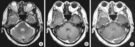
Axial T2-weighted (A) and T1-weighted (B) images show an extraaxial mass in the right sphenoid ridge area. This mass extends lateral wall of right orbit and medial aspect of right temporalis muscle. After contrast (C), strong enhancement is seen.
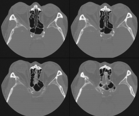
Axial computed tomography scans show irregular bony destruction with sclerotic change of the right sphenoid wing.
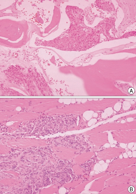
A: Photomicrographs of a section of a biopsy specimen obtained from the right sphenoid wing. Tumor cells are arranged in a sheet or lobular configuration, and individual cells have round nuclei with inconspicuous nucleoli and indistinct cytoplasmic borders. They are infiltrating the surrounding bone in a sheet-like invasion pattern (H&E×100). B: Photomicrographs of a section of a biopsy specimen obtained from the right temporalis muscle. Tumor cells are dissecting to surrounding muscle bundles and adipose tissue. Tumor cells also show also ovoid nuclei and indistinct cytoplasmic borders, forming sheets or lobules (H&E×100).
References
-
- Azar-Kia B, Sarwar M, Marc JA, Schechter MM. Intraosseous meningioma. Neuroradiology. 1974;6:246–253. - PubMed
-
- Boldrey E. The meningiomas. In: Minkler J, editor. Pathology of nervou system. Vol 2. New York: McGraw-Hill; 1971. pp. 2125–2144.
-
- Castellano F, Guidetti B, Olivecrona H. Pterional meningiomas en plaque. J Neurosurg. 1952;9:188–196. - PubMed
-
- Changhong L, Naiyin C, Yuehuan G, Lianzhong Z. Primary intraosseous meningiomas of the skull. Clin Radiol. 1997;52:546–549. - PubMed
-
- Crawford TS, Kleinschmidt-DeMasters BK, Lillehei KO. Primary intraosseous meningioma. Case report. J Neurosurg. 1995;83:912–915. - PubMed
Publication types
LinkOut - more resources
Full Text Sources
Research Materials
