

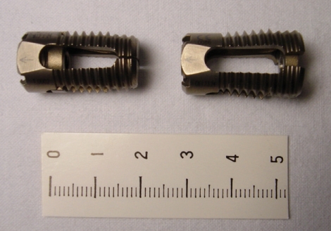
A Multi-center Clinical Study of Posterior Lumbar Interbody Fusion with the Expandable Stand-alone Cage (Tyche(R) Cage) for Degenerative Lumbar Spinal Disorders
- PMID: 19096552
- PMCID: PMC2588207
- DOI: 10.3340/jkns.2007.42.4.251
A Multi-center Clinical Study of Posterior Lumbar Interbody Fusion with the Expandable Stand-alone Cage (Tyche(R) Cage) for Degenerative Lumbar Spinal Disorders
Abstract
Objective: This multi-center clinical study was designed to determine the long-term results of patients who received a one-level posterior lumbar interbody fusion with expandable cage (Tyche(R) cage) for degenerative spinal diseases during the same period in each hospital.
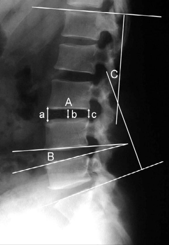
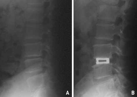
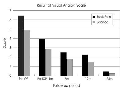
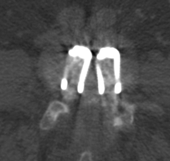
Methods: Fifty-seven patients with low back pain who had a one-level posterior lumbar interbody fusion using a newly designed expandable cage were enrolled in this study at five centers from June 2003 to December 2004 and followed up for 24 months. Pain improvement was checked with a Visual Analogue Scale (VAS) and their disability was evaluated with the Oswestry Disability Index. Radiographs were obtained before and after surgery. At the final follow-up, dynamic stability, quality of bone fusion, interveretebral disc height, and lumbar lordosis were assessed. In some cases, a lumbar computed tomography scan was also obtained.
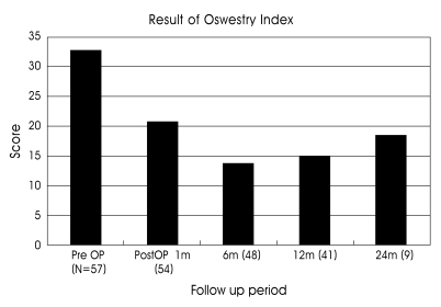
Results: The mean VAS score of back pain was improved from 6.44 points preoperatively to 0.44 at the final visit and the score of sciatica was reduced from 4.84 to 0.26. Also, the Oswestry Disability Index was improved from 32.62 points preoperatively to 18.25 at the final visit. The fusion rate was 92.5%. Intervertebral disc height, recorded as 9.94+/-2.69 mm before surgery was increased to 12.23+/-3.31 mm at postoperative 1 month and was stabilized at 11.43+/-2.23 mm on final visit. The segmental angle of lordosis was changed significantly from 3.54+/-3.70 degrees before surgery to 6.37+/-3.97 degrees by 24 months postoperative, and total lumbar lordosis was 20.37+/-11.30 degrees preoperatively and 24.71+/-11.70 degrees at 24 months postoperative.
Conclusion: There have been no special complications regarding the expandable cage during the follow-up period and the results of this study demonstrates a high fusion rate and clinical success.
Keywords: Degeneration; Expandable cage; Interbody fusion; Lumbar spine.
Figures
References
-
- Agazzi S, Reverdin A, May D. Posterior lumbar interbody fusion with cages : an independent review of 71 cases. J Neurosurg. 1999;91(Spine 2):186–192. - PubMed
-
- Attia D. Posterior lumbar interbody fusion using an original screwed titanium device : LIFEC expandable cages. A retrospective study of 48 patients with an minimal follow-up of 1 year. In: Gunzburg R, Szpalski M, editors. Lumbar spinal stenosis. Lippincott Williams and Wilkins; 2000. pp. 263–273.
-
- Barnes B, Rodts GE, McLaughlin MR, Haid RW., Jr Threaded cortical bone dowels for lumbar interbody fusion : over 1-year mean follow up in 28 patients. J Neurosurg. 2001;95(Spine 1):1–4. - PubMed
-
- Bernhardt M, Bridwell KH. Segmental analysis of the sagittal plane alignment of the normal thoracic and lumbar spines and thoracolumbar junction. Spine. 1989;14:717–721. - PubMed
-
- Brantigan JW, Steffee AD. A carbon fiber implant to aid interbody lumbar fusion. Two-year clinical results in the first 26 patients. Spine. 1993;18:2106–2117. - PubMed
LinkOut - more resources
Full Text Sources
