

Intracerebral hematoma caused by ruptured traumatic pseudoaneurysm of the middle meningeal artery : a case report
- PMID: 19096582
- PMCID: PMC2588190
- DOI: 10.3340/jkns.2007.42.5.416
Intracerebral hematoma caused by ruptured traumatic pseudoaneurysm of the middle meningeal artery : a case report
Abstract
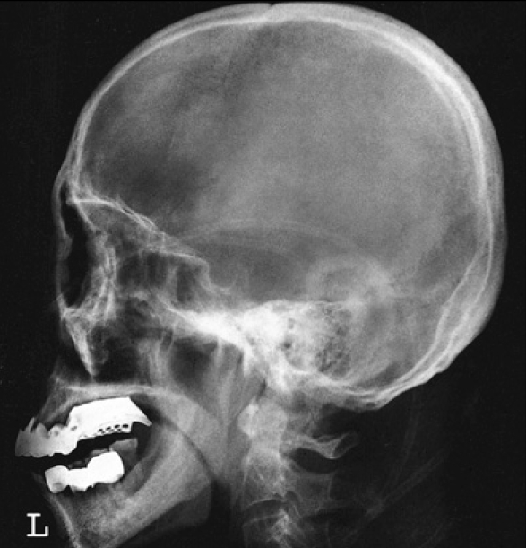
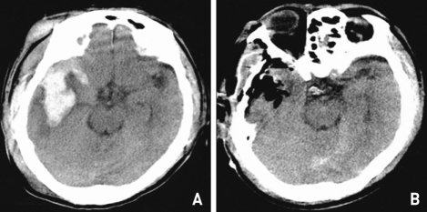
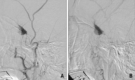
Hematomas caused by ruptured traumatic pseudoaneurysms of the middle meningeal artery (MMA) usually present with extradural hematomas, whereas intradural intraparenchymal hematomas are extremely rare. We report a case of traumatic pseudoaneurysm of the MMA giving rise to an intracerebral hematoma after head trauma. A 70-year-old man suffered a massive intracerebral temporoparietal hemorrhage after a head injury. CT angiogram of the brain revealed a large hematoma in the right middle cranial fossa extending to the right sylvian fissure. Cerebral angiogram also revealed a pseudoaneurysm of the MMA, which was successfully treated surgically. Although traumatic MMA pseudoaneurysm producing intracerebral hematoma (ICH) is rare, it should be considered as a possible cause of intracerebral hematoma.
Keywords: Intracerebral hematoma; Middle meningeal artery (MMA); Traumatic pseudoaneurysm.
Figures
References
-
- Bruneau M, Gustin T, Zekhnini K, Gilliard C. Traumatic false aneurysm of the middle meningeal artery causing an intracerebral hemorrhage : case report and literature review. Surg Neurol. 2002;57:174–178. - PubMed
-
- Garza-Mercado R, Rangel RA. Extradural hematoma associated with traumatic middle meningeal artery pseudoaneurysm. Report of two cases. Neurosurgery. 1979;5:500–503. - PubMed
-
- Higazi I, El-Banhawy A, El-Nady F. Importance of angiography in identifying false aneurysm of the middle meningeal artery as a cause of extradural hematoma. Case report. J Neurosurg. 1969;30:172–176. - PubMed
-
- Kim JH, Yim MB, Lee CY, Kim IM. Surgical management of pseudoaneurysm. J Korean Neurosurg Soc. 2001;30:307–318.
Publication types
LinkOut - more resources
Full Text Sources
