

Spinal drop metastasis from a posterior fossa choroid plexus papilloma
- PMID: 19096592
- PMCID: PMC2588175
- DOI: 10.3340/jkns.2007.42.6.475
Spinal drop metastasis from a posterior fossa choroid plexus papilloma
Abstract
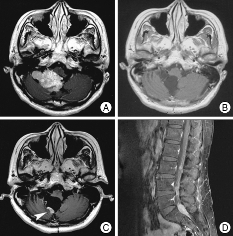
Choroid plexus papillomas (CPPs) are typically considered as benign tumors, with a favorable long-term prognosis. Drop metastasis of CPP into the spinal subarachnoid space is rare. We report a 42-year-old woman who presented with headache and back pain 6 years after removal of a posterior fossa CPP. Magnetic resonance imaging revealed mass lesions in the lumbosacral subarachnoid space and recurrent intracranial tumor. The lesions were resected and histologically diagnosed was CPP. We consider that CPP can spread via cerebrospinal fluid pathways and cause spinal drop metastasis. Therefore, it is necessary to evaluate the whole spinal axis and to perform periodic follow-up examinations in patients with CPP.
Keywords: Cerebrospinal fluid; Choroid plexus papilloma; Metastasis; Posterior fossa; Spinal.
Figures

References
-
- Enomoto H, Mizuno M, Katsumata T, Doi T. Intracranial metastasis of a choroid plexus papilloma originating in the cerebellopontine angle region : a case report. Surg Neurol. 1991;36:54–58. - PubMed
-
- Kaptanoglu E, Tun K, Celikmez RC, Ozen O, Taskin Y. Spinal drop metastasis of choroid plexus papilloma. J Clin Neurosci. 2007;14:381–383. - PubMed
-
- Leblanc R, Bekhor S, Melanson D, Carpenter S. Diffuse craniospinal seeding from a benign fourth ventricle choroid plexus papilloma. Case report. J Neurosurg. 1998;88:757–760. - PubMed
-
- McCall T, Binning M, Blumenthal DT, Jensen RL. Variations of disseminated choroid plexus papilloma : 2 case reports and a review of the literature. Surg Neurol. 2006;66:62–66. discussion 67-68. - PubMed
-
- McEvoy AW, Galloway M, Revesz T, Kitchen ND. Metastatic choroid plexus papilloma : a case report. J Neurooncol. 2002;56:241–246. - PubMed
Publication types
LinkOut - more resources
Full Text Sources
