

Distal Middle Cerebral Artery M4 Aneurysm Surgery Using Navigation-CT Angiography
- PMID: 19096593
- PMCID: PMC2588183
- DOI: 10.3340/jkns.2007.42.6.478
Distal Middle Cerebral Artery M4 Aneurysm Surgery Using Navigation-CT Angiography
Abstract
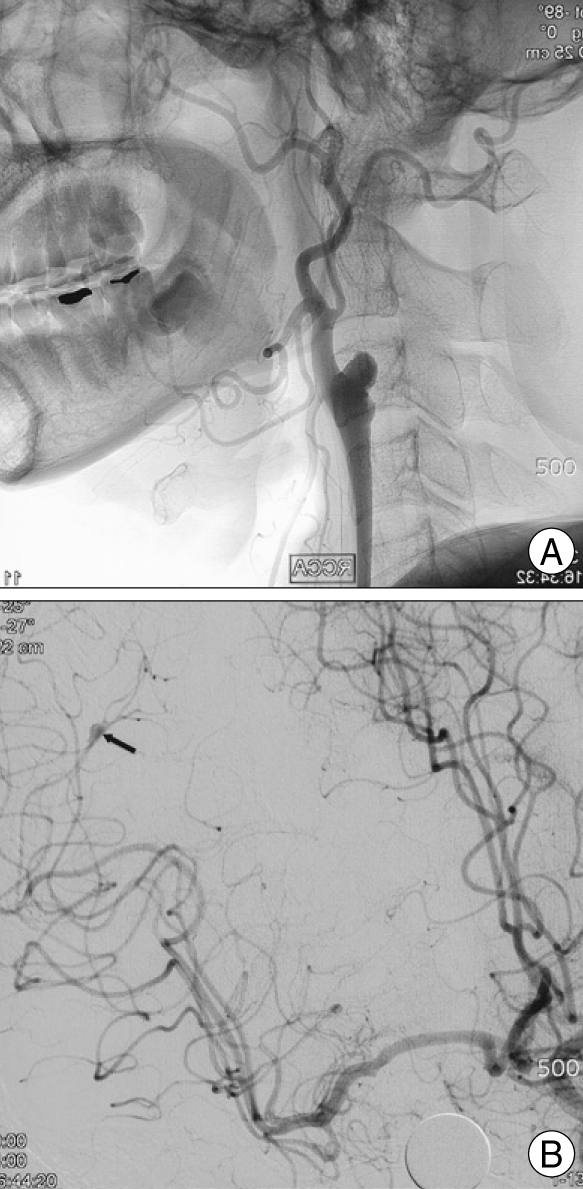
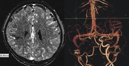
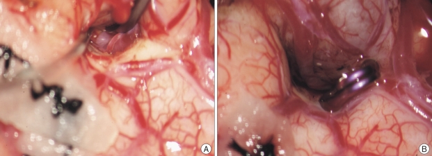
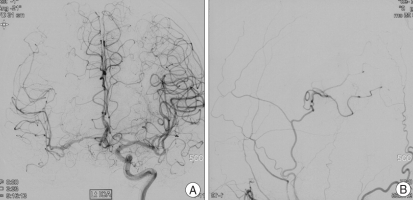
Unruptured non-traumatic dissecting aneurysm in the M4 segment of the middle cerebral artery (MCA) accompanied by complete occlusion of the ipsilateral internal cerebral artery (ICA) has never been reported. A 41-year-old man presented with an infarction manifesting as left-sided weakness and dysarthria. Magnetic resonance angiography revealed a subacute stage infarction in the right MCA territory and complete occlusion of the right ICA. Angiography demonstrated aneurysmal dilatation of the M4 segment of the right MCA. Surgery was performed to prevent hemorrhage from the aneurysm. The aneurysm was proximally clipped guided by Navigation-CT angiography and flow to the distal MCA was restored by superficial temporal artery-middle cerebral artery (STA-MCA) anastomosis. We report this rare case with literature review.
Keywords: Dissecting aneurysm; Middle cerebral artery aneurysm; Navigation.
Figures
References
-
- Ahn JY, Han IB, Joo JY. Aneurysm in the penetrating artery of the distal middle cerebral artery presenting as intracerebral haemorrhage. Acta Neurochir (Wien) 2005;147:1287–1290. discussion 1290. - PubMed
-
- Lee KS, Liu SS, Spetzler RF. Intracranial mycotic aneurysm in an infant : report of a case. Neurosurgery. 1990;26:129–133. - PubMed
-
- Rinne J, Hernesniemi J, Niskanen M. Analysis of 561 patients with 690 middle cerebral artery aneurysms : anatomic and clinical features as correlated to management outcome. Neurosurgery. 1996;38:2–11. - PubMed
-
- Sakamoto S, Ikawa F, Kawamoto H. Acute surgery for ruptured dissecting aneurysm of the M3 portion of the middle cerebral artery. Neurol Med Chir (Tokyo) 2003;43:188–191. - PubMed
-
- Schmid-Elsaesser R, Muacevic A, Holtmannspotter M. Neuronavigation based on CT angiography for surgery of intracranial aneurysms : primary experience with unruptured aneurysms. Minim Invasive Neurosurg. 2003;46:269–277. - PubMed
Publication types
LinkOut - more resources
Full Text Sources
Miscellaneous
