Pituitary Apoplexy due to Pituitary Adenoma Infarction
- PMID: 19096606
- PMCID: PMC2588219
- DOI: 10.3340/jkns.2008.43.5.246
Pituitary Apoplexy due to Pituitary Adenoma Infarction
Abstract
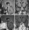
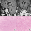
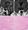
Cause of pituitary apoplexy has been known as hemorrhage, hemorrhagic infarction or infarction of pituitary adenoma or adjacent tissues of pituitary gland. However, pituitary apoplexy caused by pure infarction of pituitary adenoma has been rarely reported. Here, we present the two cases pituitary apoplexies caused by pituitary adenoma infarction that were confirmed by transsphenoidal approach (TSA) and pathologic reports. Pathologic report of first case revealed total tumor infarction of a nonfunctioning pituitary macroadenoma and second case partial tumor infarction of ACTH secreting pituitary macroadenoma. Patients with pituitary apoplexy which was caused by pituitary adenoma infarction unrelated to hemorrhage or hemorrhagic infarction showed good response to TSA treatment. Further study on the predisposing factors of pituitary apoplexy and the mechanism of infarction in pituitary adenoma is necessary.
Keywords: Pituitary adenoma infarction; Pituitary apoplexy.
Figures


References
-
- Alarifi A, Alzahrani AS, Salam SA, Ahmed M, Kanaan I. Repeated remissions of cushing disease due to recurrent infarctions of an ACTH producing pituitary macroadenoma. Pituitary. 2005;8:81–87. - PubMed
-
- Brougham M, Heusner AP, Adams RD. Acute degeneration changes in adenomas of pituitary body with special reference to pituitary apoplexy. J Neurosurg. 1950;7:421–439. - PubMed
-
- Cardoso ER, Peterson EW. Pituitary apoplexy : a review. Neurosurgery. 1984;14:363–373. - PubMed
-
- Elsasser Imboden PN, De Tribolet N, Lobrinus A, Gaillard RC, Portmann L, Pralong F, et al. Apoplexy in pituitary macroadenoma : eight patients presenting in 12 months. Medicine (Baltimore) 2005;84:188–196. - PubMed
Publication types
LinkOut - more resources
Full Text Sources
Research Materials

