

Fusiform intracanalicular ophthalmic artery aneurysm; case report and review of literature
- PMID: 19096656
- PMCID: PMC2588290
- DOI: 10.3340/jkns.2008.44.1.43
Fusiform intracanalicular ophthalmic artery aneurysm; case report and review of literature
Abstract
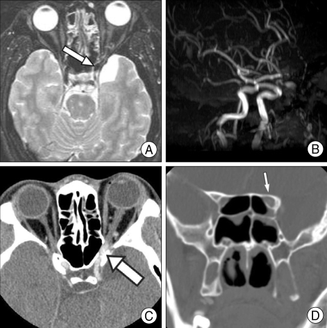
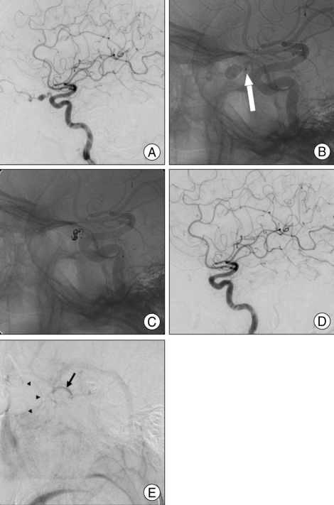
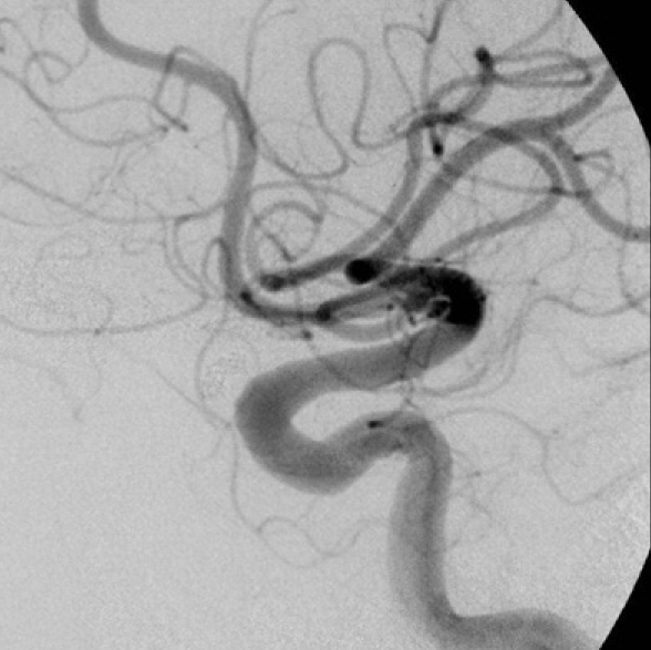
A 35-year-old man's vision had progressively deteriorated over a 3-month period. His left visual acuity was 5/20. Enhanced orbital computed tomographic (CT) scans revealed a fusiform dilatation of the ophthalmic artery in the left optic canal. Cerebral Angiography revealed a fusiform aneurysm on the left ophthalmic artery in the optic canal, measuring 6.2 x 4.6 mm in size. Four days after admission, visual acuity dropped to hand-motion. Endovascular treatment was chosen and a microcatheter was guided into the proximal segment of the ophthalmic artery. Using 4 detachable coils, parent artery occlusion was done. Three months after the intervention, the visual acuity in his left eye improved to 20/20. Dramatic recovery of visual acuity is exceptional with an ophthalmic artery trunk aneurysm. When an occlusion of the proximal ophthalmic artery is the only treatment option in such a situation, the endovascular occlusion of the proximal ophthalmic artery is quite feasible in the sense that it does not require any optic nerve manipulation.
Keywords: Detachable coil; Fusiform aneurysm; Intracanalicular portion; Ophthalmic artery trunk aneurysm.
Figures
References
-
- Beiran I, Dori D, Pikkel J, Goldsher D, Miller B. Recurrent retinal artery obstruction as a presenting symptom of ophthalmic artery aneurysm : a case report. Graefes Arch Clin Exp Ophthalmol. 1995;233:444–447. - PubMed
-
- Dehdashti AR, Safran AB, Martin JB, Rufenacht DA, de Tribolet N. Intraorbital ophthalmic artery aneurysm associated with basilar tip saccular aneurysm. Neuroradiology. 2002;44:600–603. - PubMed
-
- Drake CG, Vanderlinden RG, Amacher AL. Carotid-ophthalmic aneurysms. J Neurosurg. 1968;29:24–31. - PubMed
-
- Ernemann U, Freudenstein D, Pitz S, Naegele T. Intraorbital aneurysm of the ophthalmic artery : a rare cause of apex orbitae compression syndrome. Graefes Arch Clin Exp Ophthalmol. 2002;240:575–577. - PubMed
-
- Jain KK. Saccular aneurysm of the ophthalmic artery. Am J Ophthalmol. 1970;69:997–998. - PubMed
Publication types
LinkOut - more resources
Full Text Sources
