Review
. 2009:(190):403-18.
doi: 10.1007/978-3-540-79885-9_20.

Aquaporin-1 gene transfer to correct radiation-induced salivary hypofunction

Affiliations
Review

Aquaporin-1 gene transfer to correct radiation-induced salivary hypofunction

Bruce J Baum et al. Handb Exp Pharmacol. 2009.

Abstract

Irradiation damage to salivary glands is a common iatrogenic consequence of treatment for head and neck cancers. The subsequent lack of saliva production leads to many functional and quality-of-life problems for affected patients and there is no effective conventional therapy. To address this problem, we developed an in vivo gene therapy strategy involving viral vector-mediated transfer of the aquaporin-1 cDNA to irradiation-damaged glands and successfully tested it in two pre-clinical models (irradiated rats and miniature pigs), as well as demonstrated its safety in a large toxicology and biodistribution study. Thereafter, a clinical research protocol was developed that has received approval from all required authorities in the United States. Patients are currently being enrolled in this study.


Figures

Fig. 1
Fig. 1
Schematic diagram of the hypothesized mechanism for fluid secretion following AdhAQP1-mediated gene transfer to duct cells in an irradiated salivary gland. The surviving duct cell is presented in a simplified form, based on our understanding ∼1991. The duct lumen is to the left and the interstitium is to the right. The three apical ion pathways with Xs will be inoperable in the absence of an isotonic primary secretion, which normally is made by acinar cells. We have hypothesized that duct cells could generate a KHCO3 gradient (lumen > interstitium) enabling fluid to flow into the duct lumen following expression of the transgene hAQP1. This figure is modified from Vitolo and Baum (2002), and is based on the experiments presented in Delporte et al. (1997)
Fig. 2
Fig. 2
Schematic diagram of AdhAQP1. ITR inverted terminal repeat; Pcmv cytomegalovirus promoter/enhancer; hAQP1 human aquaporin-1 cDNA; SV40 polyA simian virus 40 polyadenylation signal; ΔE1 deletion of adenoviral E1 sequences; E2 adenoviral E2 genes; pΔE3 partial deletion/modification of adenoviral E3 sequences; E4 adenoviral E4 genes
Fig. 3
Fig. 3
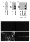
Human aquaporin-1 expression in epithelial cells in vitro. Upper panel: (a) Northern blot using RNA from 293 cells transduced with either AdhAQP1 or a control vector, Adα1AT. (b) Western blot of crude membranes from 293 cells transduced with AdhAQP1 or the control vector. (c) Western blot of crude membranes from MDCK and SMIE cells transduced with AdhAQP1 or the control vector. Note that in the Western blots the monomeric non-glycosylated hAQP1 protein migrates at ∼28kDa, while multiple glycosylated forms are seen at slightly higher molecular weights. This figure originally was published as Fig. 1 in (Delporte et al. 1997). Lower panel: Localization of transgenic hAQP1 expressed in MDCK cells. Confluent MDCK cells were grown on filters and transduced for 24 h with either Adα1AT (a) and (b) or AdhAQP1 (c) and (d). Cell layers were then examined by confocal microscopy after immunofluorescent staining with an antibody to hAQP1. (a) and (c) are in the x–y plane, while (b) and (d) are in the x–z plane. This figure originally was published as Fig. 2 in (Delporte et al. 1997)
Fig. 4
Fig. 4
Net fluid secretion rate of MDCK cells with and without transduction by AdhAQP1. Cells were either untreated (control, Cont), or transduced with either AdhAQP1 or a control vector, Adα1AT, and water flow measured in response to an apical (400 mosm) >basal (300 mosm) osmotic gradient. The results are expressed as water flow in microliters of fluid per cm2 per hour and are mean values ± SEMs. This figure originally was published as Fig. 3 in (Delporte et al. 1997)
Fig. 5
Fig. 5
Time course of fluid movement across SMIE cell monolayers. SMIE cells were transduced with AdhAQP1 (MOI = 5) or not, and after 24 h fluid movement was measured in response to an osmotic gradient as shown in Fig. 4. The results are expressed as water flow in microliters of fluid per cm2 per hour and are mean values ± SEMs. This figure originally was published as Fig. 2 in (Delporte et al. 1998)
Fig. 6
Fig. 6
Effect of vector concentration on fluid flow across SMIE cells. After reaching confluence, SMIE cell monolayers were transduced at the indicated MOI and fluid movement then measured for 15 min. Thereafter, crude membranes from each cell monolayer were prepared, electrophoresed and subjected to Western blotting with antibody to hAQP1. The films were scanned with a laser densitometer to quantify the amount of hAQP1 expressed (indicated in arbitrary units). This figure originally was published as Fig. 4 in (Delporte et al. 1998)
Fig. 7
Fig. 7
(a) Time line of initial study of AdhAQP1 efficacy in irradiated rats. Rats were irradiated on day zero with either 17.5 Gy or 21 Gy, or sham-irradiated. After 90 (17.5 Gy) or 120 (21 Gy) days, AdhAQP1 or a control vector (Addl312) was administered to both submandibular glands and 3 days later saliva was collected. (b) Function of transgenic hAQP1 in vivo. Salivary flow rates obtained in animals irradiated (17.5 Gy) or not, and administered either AdhAQP1 or Addl312. Rats administered Addl312 are shown in the hatched bars, while rats administered AdhAQP1 are shown in the black bars. The results are expressed as saliva secretion in microliters per 100 g body weight per 15 min and are mean values ± SEMs. This figure originally was published as Fig. 4b in (Delporte et al. 1997)
Fig. 8
Fig. 8
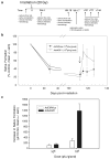
The effect of AdhAQP1 vector administration on parotid salivary secretion in irradiated miniature pigs. (a) Time line of study. Animals (three separate cohorts) in these experiments were followed longitudinally. Either AdhAQP1, a control vector (AdCMVluc, encoding luciferase), or buffer was given as indicated. (b) Pattern of parotid salivary flow following irradiation and vector administration. Parotid salivary flow rates (in μl per 10 min; average of two measurements) prior to irradiation were normalized to 100% and data at other time-points are shown as a percentage of that initial value. The arrow indicates the time point when vectors were administered. (c) Effect of AdhAQP1 dose on parotid saliva secretion. All data shown are mean values ± SEMs. This figure originally was published as part of Fig. 1 in (Shan et al. 2005)
Fig. 9
Fig. 9
Representative clinical chemistry results seen with female rats treated, or not, with various doses of AdhAQP1. ALT alanine aminotransferase (∼liver damage); CPK creatine phosphokinase (∼heart damage); BUN blood urea nitrogen (∼kidney function); LDH lactate dehydrogenase (indicates general tissue damage). Data shown are mean values ± SD. Dosage groups are indicated (vector genomes of AdhAQP1 administered). This figure was published originally as Fig. 4 in (Zheng et al. 2006)
Fig. 10
Fig. 10
Animal body weights during AdhAQP1 toxicology study. Left panel: Mean body weight in grams is depicted for all four groups of male rats over the time course of this study. Right panel: Mean body weight in grams is depicted for all four groups of female rats over the time course of this study. The animal study groups are as indicated (doses in vector genomes of AdhAQP1). This figure was published originally as Fig. 1 in (Zheng et al. 2006)

Similar articles

Cited by

References

    1. Adesanya MR, Redman RS, Baum BJ, et al. Immediate inflammatory responses to adenovirus-mediated gene transfer in rat salivary glands. Hum Gene Ther. 1996;7:1085–1093. - PubMed
    1. Baum BJ. Principles of saliva secretion. Ann N Y Acad Sci. 1993;694:17–23. - PubMed
    1. Baum BJ, Wang S, Cukierman E, et al. Re-engineering the functions of a terminally differentiated epithelial cell in vivo. Ann N Y Acad Sci. 1999;875:294–300. - PubMed
    1. Baum BJ, Wellner RB, Zheng C. Gene transfer to salivary glands. Int Rev Cytol. 2002;213:93–146. - PubMed
    1. Delporte C, O'Connell BC, He X, et al. Increased fluid secretion after adenoviral-mediated transfer of the aquaporin-1 cDNA to irradiated rat salivary glands. Proc Natl Acad Sci U S A. 1997;94:3268–3273. - PMC - PubMed

Publication types

MeSH terms