. 2009 Mar;467(3):739-46.
doi: 10.1007/s11999-008-0659-8. Epub 2008 Dec 19.

Arthroscopic femoroplasty in the management of cam-type femoroacetabular impingement

Affiliations

Arthroscopic femoroplasty in the management of cam-type femoroacetabular impingement

J W Thomas Byrd et al. Clin Orthop Relat Res. 2009 Mar.

Abstract

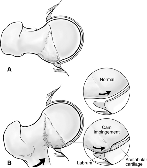
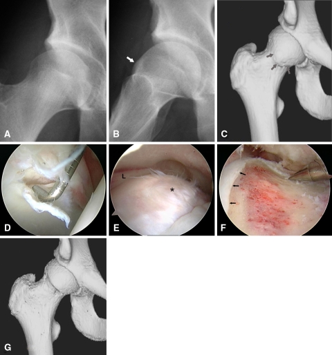
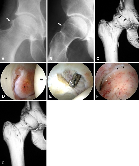
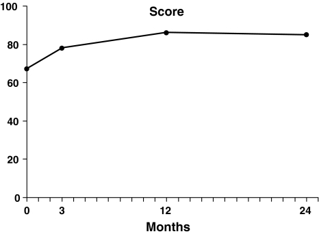
Cam-type femoroacetabular impingement is a recognized cause of intraarticular pathology and secondary osteoarthritis in young adults. Arthroscopy is reportedly useful to treat selected hip abnormalities and has been proposed as a method of correcting underlying impingement. We report the outcomes of arthroscopic management of cam-type femoroacetabular impingement. We prospectively assessed all 200 patients (207 hips) who underwent arthroscopic correction of cam impingement from December 2003 to October 2007, using a modified Harris hip score. The minimum followup was 12 months (mean, 16 months; range, 12-24 months); no patients were lost to followup. The average age was 33 years with 138 men and 62 women. One hundred and fifty-eight patients (163 hips) underwent correction of cam impingement (femoroplasty) alone while 42 patients (44 hips) underwent concomitant correction of pincer impingement. The average increase in Harris hip score was 20 points; 0.5% converted to THA. We had a 1.5% complication rate. The short-term outcomes of arthroscopic treatment of cam-type femoroacetabular impingement are comparable to published reports for open methods with the advantage of a less invasive approach.


Figures

Fig. 1A–B
Fig. 1A–B
Diagrams illustrate (A) cam morphology created by bony prominence at the anterolateral head-neck junction, resulting in a nonspherical femoral head; and (B) dynamics of cam impingement that occur with hip flexion. Compared to normal, the cam lesion glides underneath the labrum engaging the edge of the articular cartilage and results in progressive delamination. Initially the labrum is relatively preserved, but secondary failure occurs over time. (Reprinted with permission from J. W. Thomas Byrd, MD.)
Fig. 2A–G
Fig. 2A–G
Images illustrate the case of a 20-year-old hockey player with a 4-year history of right hip pain. (A) An anteroposterior radiograph is unremarkable. (B) A frog lateral radiograph demonstrates a morphologic variant with bony buildup at the anterior femoral head-neck junction (arrow) characteristic of cam impingement. (C) A 3-D CT scan further defines the extent of the bony lesion (arrows). (D) Viewing from the anterolateral portal, the probe introduced anteriorly displaces an area of articular delamination from the anterolateral acetabulum characteristic of the peel back phenomenon created by the bony lesion shearing the articular surface during hip flexion. (E) Viewing from the peripheral compartment, the bony lesion is identified (*) immediately below the free edge of the acetabular labrum (L). (F) The lesion has been excised, recreating the normal concave relationship of the femoral head-neck junction immediately adjacent to the articular surface (arrows). Posteriorly, resection is limited to the midportion of the lateral neck to avoid compromising blood supply to the femoral head from the lateral retinacular vessels. (G) A postoperative 3-D CT scan illustrates the extent of bony resection. (Reprinted with permission from J. W. Thomas Byrd, MD.)
Fig. 3A–G
Fig. 3A–G
Images illustrate the case of a 38-year-old woman with progressive pain and loss of motion of the right hip. (A) An anteroposterior radiograph demonstrates acquired bony buildup/osteophyte formation on the lateral femoral head (arrow). (B) A frog lateral radiograph further defines the bony buildup on the anterior femoral head (arrow). (C) A 3-D CT scan further defines the femoral head osteophyte (*) and the anterior acetabular lesion (arrows). (D) Arthroscopy of the central weightbearing surface of the joint demonstrates good articular preservation of both the acetabulum (A) and femoral head (FH) with some reactive synovitis within the fossa (S). (E) The anterior acetabular osteophyte is excised. (F) Viewing peripherally, the femoral head has been recontoured showing the edge of the femoral articular surface (white arrows) and the labrum (black arrows). (G) A postoperative 3-D CT scan demonstrates the extent of bony recontouring of both the acetabulum and femoral head. (Reprinted with permission from J. W. Thomas Byrd, MD.)
Fig. 4
Fig. 4
Change in modified Harris hip scores over time. Continued improvement can be seen throughout the first year, with results maintained in those with 2-year followup. (Reprinted with permission from J. W. Thomas Byrd, MD.)

Similar articles

Cited by

References

    1. {'text': '', 'ref_index': 1, 'ids': [{'type': 'DOI', 'value': '10.2106/JBJS.F.00681', 'is_inner': False, 'url': 'https://doi.org/10.2106/jbjs.f.00681'}, {'type': 'PubMed', 'value': '17403799', 'is_inner': True, 'url': 'https://pubmed.ncbi.nlm.nih.gov/17403799/'}]}
    2. Beaulé PE, Le Duff MJ, Zaragoza E. Quality of life following femoral head-neck osteochondroplasty for femoroacetabular impingement. J Bone Joint Surg Am. 2007;89:773–779. - PubMed
    1. {'text': '', 'ref_index': 1, 'ids': [{'type': 'DOI', 'value': '10.1097/00003086-200401000-00012', 'is_inner': False, 'url': 'https://doi.org/10.1097/00003086-200401000-00012'}, {'type': 'PubMed', 'value': '15043095', 'is_inner': True, 'url': 'https://pubmed.ncbi.nlm.nih.gov/15043095/'}]}
    2. Beck M, Leunig M, Parvizi J, Boutier V, Wyss D, Ganz R. Anterior femoroacetabular impingement: part II. Midterm results of surgical treatment. Clin Orthop Relat Res. 2004;418:67–73. - PubMed
    1. {'text': '', 'ref_index': 1, 'ids': [{'type': 'PubMed', 'value': '8086020', 'is_inner': True, 'url': 'https://pubmed.ncbi.nlm.nih.gov/8086020/'}]}
    2. Byrd JW. Hip arthroscopy utilizing the supine position. Arthroscopy. 1994;10:275–280. - PubMed
    1. {'text': '', 'ref_index': 1, 'ids': [{'type': 'DOI', 'value': '10.1053/j.oto.2004.04.010', 'is_inner': False, 'url': 'https://doi.org/10.1053/j.oto.2004.04.010'}]}
    2. Byrd JW. Hip arthroscopy: evolving frontiers. Op Tech Orthop. 2004;14:58–67.
    1. {'text': '', 'ref_index': 1, 'ids': [{'type': 'PubMed', 'value': '10976117', 'is_inner': True, 'url': 'https://pubmed.ncbi.nlm.nih.gov/10976117/'}]}
    2. Byrd JW, Jones KS. Prospective analysis of hip arthroscopy with two year follow up. Arthroscopy. 2000;16:578–587. - PubMed

MeSH terms