

doi: 10.1002/anie.200804107.
Ammosamides A and B target myosin
Affiliations
- PMID: 19097126
- PMCID: PMC2820877
- DOI: 10.1002/anie.200804107
Item in Clipboard
Ammosamides A and B target myosin
Angew Chem Int Ed Engl.
2009.
No abstract available
Figures
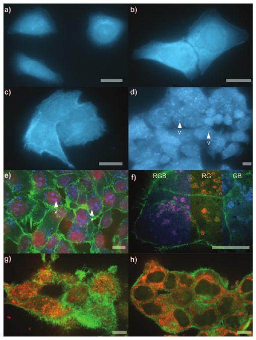
Cellular phenotype. a–c) Images from 106 cells incubated with 1 mL of 50 μm probe 4 in DMEM for 15 min. a) HeLa cells. b) HCT-116 cells. c) PC-3 cells. d) HeLa cells in (a) after incubation at 37°C with 5% CO2 for 12 h. e) Three-colored confocal micrographs of the cells in (d) after fixation and staining of the nucleus with Syto608[9a] (R), actin with FITC-phalloidin,[9b] (G) and probe 4 (B). f) Cells in (d) after staining the lysosomes with LysoTracker Red DND-99[9c] (R), actin with FITC-phalloidin[9b] (G) and probe 4 (B). Color mixing of R and B channels overlap to form magenta as depicted by three color mixing (RGB). Individual R and B channels are shown by RG and GB composites. g,h) HCT-116 cells treated for 5 h with 1 mL of 50 μm probe 4 in DMEM per 106 cells, fixed, and then stained. Microtubules were stained with BODIPY 564/570 paclitaxel[9d] (R) and actin with FITC-phalloidin (G). Colors are denoted as (R)=red, (B)=blue, (G)=green. Bar denotes 10 μm.
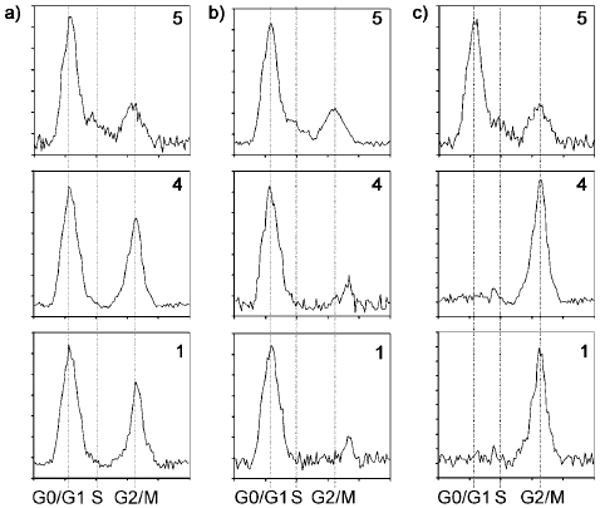
Cell cycle response. HCT-116 cells were treated with either 50 μm control dye 5 (normal cell proliferation), probe 4 or ammosamide A (1) in DMEM per 106 cells. a) Unsynchronized HCT-116 cells. b) HCT-116 cells synchronized and treated at G0 and incubated for 12 h. c) HCT-116 cells synchronized and treated at S and incubated for 12 h.
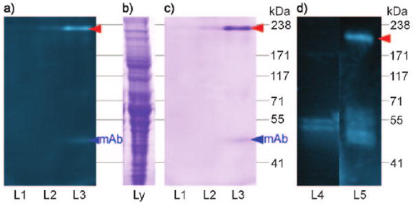
Co-immunoprecipitation (co-IP) studies. a) A 3–8% Trisacetate SDS-PAGE gel depicting fluorescent bands arising from the co-IP of lysate from 108 HCT-116 cells treated with probe 4 and Affigel Hz resin containing 12.5 mg mL−1 of XRI-TF35 mAb. After incubation for 12 h and multiple washings with PBS pH 7.2 at 4°C, the bound protein was eluted from XRI-TF35-Affigel Hz resin with 0.1 m Tris-Cl pH 6.8 (L1), 5 μm 3 in 0.1 m Tris-Cl pH 6.8 (L2), or 50 μm 3 in 0.1 m Tris-Cl pH 6.8 (L3) at 23 °C. b) HCT-116 lysate stained with GelCode blue. c) GelCode blue staining of the gel in (a). d) A 4% Tris-glycine SDS-PAGE gel depicting fluorescent bands from the co-IP of a 50 μg mL−1 sample of rabbit skeletal myosin that was incubated with 10 μm control 5 in PBS pH 7.2 (L4) or 10 μm probe 4 in PBS pH 7.2 (L5) and Affigel Hz resin containing 12.5 mg mL−1 of XRI-TF35 mAb. The bands in L4 and L5 were obtained after multiple washings of the resin with PBS pH 7.2 at 4°C and subsequent elution of the bound protein with 50 μm 3 in PBS pH 7.2 at 23 °C. Red arrow denotes bands of interest.
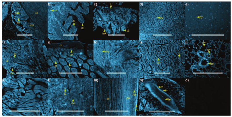
Histological analysis. A microarray containing select Mus musculus tissues[13] was treated for 4 h with 2 mL of 1 μm probe 4 in PBS pH 7.2. Images from each tissue section were collected using an identical exposure, thereby allowing the intensity to be compared between each image. Tissues stained with probe 4: a) adrenal gland, b) bladder, c) bone marrow, d) cerebellum, e) cerebral cortex, f) breast, g) intestine, h) heart, i) kidney, j) lung, k) skeletal muscle, l) pancreas, m) skin, and n) spleen. o) Control experimentation was conducted in parallel by treating the tissue microarray under the identical conditions used to collect the images in (a–m) with 1 μm control dye 5 in PBS pH 7.2. For all panels, the background from 5 was not visible under the exposure time used for image collection, as demonstrated by the uptake control probe 5 in heart tissue. Anatomical denominations: a=adipose cells, ac=acini, av=alveoli, br=bronchiole, c=capillaries, cc=chrimaffin cells, cp=crypts, ct=connecting tubules, cx=cortex, de=dermis, du=ducts, e=epithelial cells, ed=epidermis, ei=erythroid island, iL=islet of Langerhans, lp=lamina propria, m=muscularis, md=medulla, mk=megakaryocyte, my=myocytes, pv=pulminary vetricle, py=pyramidal cells, s=sinusoids, sc=subcutis, sg=sebaceous glands, sr=serosa, tb=trabecular bone, te=transitional epithelium, uc=umbrella cells, v=villus. Bars denote 1 mm.
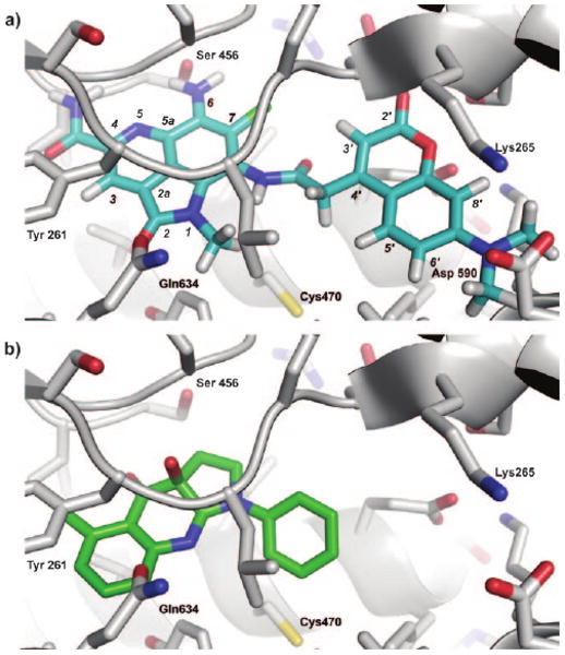
Docking studies. a) A depiction of ammosamide probe 4 (blue) docked in Dictyostelium discoideum myosin II. b) The binding of (S)-(−)-blebbistatin (green) within the same pocket of Dictyostelium discoideum myosin II. Docking was conducted with Autodock 3 using the coordinates from PDB accession number 3bz7.[16]
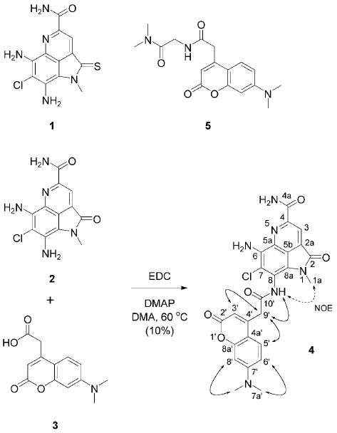
Structures of ammosamide A (1), ammosamide B (2), 7-dimethylaminocoumarin-4-acetic acid (3), ammosamide B probe 4 and control dye 5. Probe 4 was prepared by coupling 2 and 3. ROESY (bold arrows) and NOESY correlations (dotted arrows) used in the elucidation of the structure of probe 4 are shown. EDC=N′-(3-dimethylaminopropyl)-N-ethylcarbodiimide; DMAP=4-dimethylaminopyridine; DMA=N,N-dimethylacetamide.
References
Publication types
MeSH terms
Substances
Grants and funding
LinkOut - more resources
Full Text Sources
Other Literature Sources
