. 2010 May;31(5):796-804.
doi: 10.1016/j.neurobiolaging.2008.11.001. Epub 2008 Dec 20.

Absence of alpha-synuclein affects dopamine metabolism and synaptic markers in the striatum of aging mice

Affiliations

Absence of alpha-synuclein affects dopamine metabolism and synaptic markers in the striatum of aging mice

Abdelmojib Al-Wandi et al. Neurobiol Aging. 2010 May.

Abstract

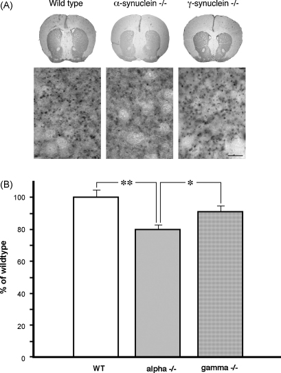
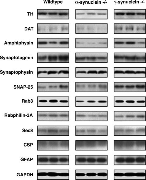
Despite numerous evidences for neurotoxicity of overexpressed alpha-synuclein, a protective function was suggested for endogenous alpha-synuclein and other members of the synuclein family. This protective role is most important for and evident in presynaptic terminals, where synucleins are normally accumulated. However, mice lacking synucleins display no adverse phenotype. In particular, no significant changes in striatal dopamine metabolism and only subtle deficit of dopaminergic neurons in the substantia nigra were found in juvenile or adult mice. To assess whether aging and synuclein deficiency may have additive detrimental effect on the nigrostriatal system, we studied dopaminergic neurons of the substantia nigra and their striatal synapses in 24-26-month-old alpha-synuclein and gamma-synuclein null mutant mice. Significant approximately 36% reduction of the striatal dopamine was found in aging alpha-synuclein, but not gamma-synuclein null mutant mice when compared to age-matching wild type mice. This was accompanied by the reduction of TH-positive fibers in the striatum and decrease of striatal levels of TH and DAT. However, no progressive loss of TH-positive neurons was revealed in the substantia nigra of synuclein-deficient aging animals. Our results are consistent with a hypothesis that alpha-synuclein is important for normal function and integrity of synapses, and suggest that in the aging nervous system dysfunction of this protein could become a predisposition factor for the development of nigrostriatal pathology.


Figures

Fig. 1
Fig. 1
Dopamine and its metabolite levels in striatum of 2-year-old wild type and synuclein null mutant mice. Striatal concentrations (ng/mg protein) of dopamine (DA), 3,4-dihydroxyphenylacetic acid (DOPAC) and homovanillic acid (HVA) in mutant animals were normalized to corresponding mean values for wild type animals (100%) in each experiment. Means ± S.E.M. for 16 wild type (WT), 11 α-synuclein null mutant (alpha−/−) and 10 γ-synuclein null mutant (gamma−/−) animals from two separate experimental cohorts are shown. Statistical analysis revealed significant decrease of striatal DA in α-synuclein null mutant mice (*, p < 0.05, one-way ANOVA with post hoc Fisher’s protected t-test) but no differences for other neurochemicals and genotypes (p > 0.05).
Fig. 2
Fig. 2
The number of dopaminergic neurons in the substantia nigra and ventral tegmental area of 2-year-old wild type and synuclein null mutant mice and their performance in accelerated rotarod test. Bar chart shows means ± S.E.M. of total number of TH-positive neurons in SNpc (A) and VTA (B). Neurons were counted separately in left and right SNpc and VTA of 9 wild type (WT), 9 α-synuclein null mutant (alpha−/−) and 10 γ-synuclein null mutant (gamma−/−) animals. Statistic analysis revealed significantly reduced number of neurons in SNpc for both types of mutant mice when compared to wild type mice (*, p < 0.01, one-way ANOVA with post hoc Fisher’s protected t-test) and no difference in number of VTA neurons between all three groups (p > 0.05). Bar chat in panel C shows results of an accelerated rotarod test that demonstrated no significant difference (p > 0.05) in latency to fall between three groups of 2-year-old mice.
Fig. 3
Fig. 3
Immunohistochemical detection of tyrosine hydroxylase in the striatum of 2-year-old wild type and synuclein null mutant mice. Representative microphotographs of coronal sections of wild type, α-synuclein and γ-synuclein null mutant mouse brains at the Bregma 1.18 mm level stained with antibody against TH (top panels) and high magnification images that reveal TH-positive fibers in the dorsal striatum (bottom panes, scale bar = 10 μm) are shown (A). Bar chart shows means ± S.E.M. of TH staining densities (B). One-way ANOVA with post hoc Fisher’s protected t-test demonstrated that the density is significantly lower in the striatum of α-synuclein (alpha−/−) null mutant mice when compared to wild type (WT) mice (**p < 0.01) or γ-synuclein (gamma−/−) null mutant mice (*p < 0.05).
Fig. 4
Fig. 4
Synaptic marker proteins in the striatum of 2-year-old mice. Representative Western blots of striatal proteins extracted from wild type, α-synuclein and γ-synuclein null mutant mice are shown. Samples extracted from each dissected striatum individually were first normalized using anti-β-actin antibody as described in Section 2. Equal amount of total protein was loaded on each lane and probed with antibody against proteins shown left to each horizontal panel.
Fig. 5
Fig. 5
β-synuclein in the striatum of 2-year-old mice. (A) Bar chart shows means ± S.E.M. of β-synuclein/GAPDH band density ratios normalized to the mean ratio for wild type animal samples as 100%. Individual striatum samples from 7 to 9 animals per genotype were analysed by Western blotting. No significant differences were found between groups (p > 0.05). (B) A representative Western blot shows analysis of mixtures of the equal amounts of total protein samples from three striata for each genotype. The filters were probed simultaneously with anti-β-synuclein and anti-GAPDH antibody.

Similar articles

Cited by

References

    1. Abeliovich A., Schmitz Y., Farinas I., Choi-Lundberg D., Ho W.H., Castillo P.E., Shinsky N., Verdugo J.M., Armanini M., Ryan A., Hynes M., Phillips H., Sulzer D., Rosenthal A. Mice lacking alpha-synuclein display functional deficits in the nigrostriatal dopamine system. Neuron. 2000;25:239–252. - PubMed
    1. Abercrombie M. Estimation of nicear population from microtome sections. Anat. Rec. 1946;94:239–247. - PubMed
    1. Altar C.A., Marien M.R., Marshall J.F. Time course of adaptations in dopamine biosynthesis, metabolism, and release following nigrostriatal lesions: implications for behavioral recovery from brain injury. J. Neurochem. 1987;48:390–399. - PubMed
    1. Alvarez-Fischer D., Henze C., Strenzke C., Westrich J., Ferger B., Hoglinger G.U., Oertel W.H., Hartmann A. Characterization of the striatal 6-OHDA model of Parkinson’s disease in wild type and alpha-synuclein-deleted mice. Exp. Neurol. 2008;210:182–193. - PubMed
    1. Benno R.H., Tucker L.W., Joh T.H., Reis D.J. Quantitative immunocytochemistry of tyrosine hydroxylase in rat brain. I. Development of a computer assisted method using the peroxidase-antiperoxidase technique. Brain Res. 1982;246:225–236. - PubMed

Publication types

MeSH terms