

Evaluation of endosonography as a new diagnostic tool for feline pancreatitis
- PMID: 19097924
- PMCID: PMC10832838
- DOI: 10.1016/j.jfms.2008.11.007
Evaluation of endosonography as a new diagnostic tool for feline pancreatitis
Abstract
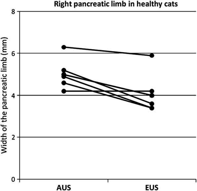
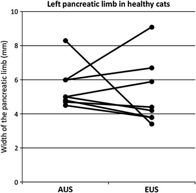
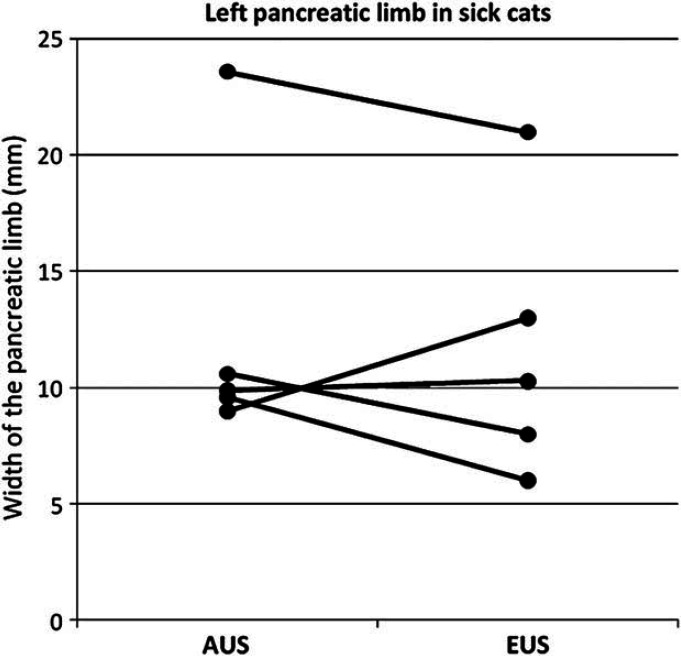
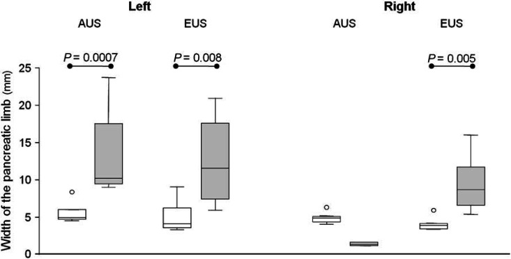
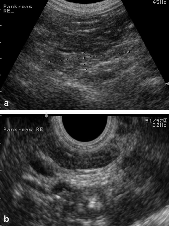
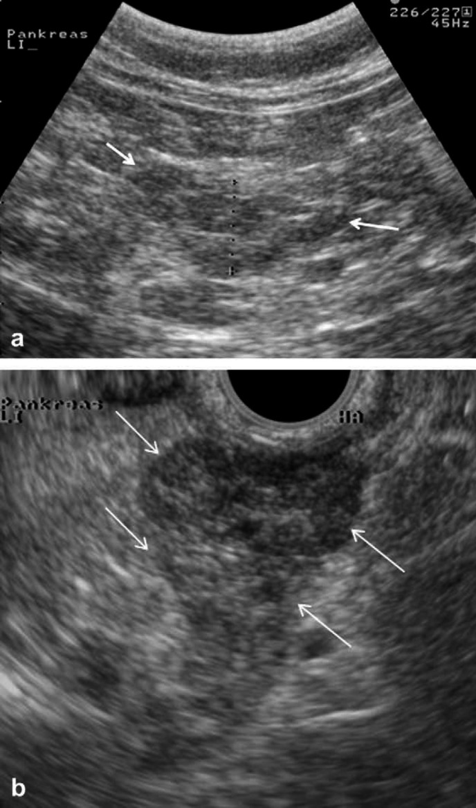
The purpose of this study was to evaluate endosonography (EUS) as a potential diagnostic tool for feline pancreatitis. Eleven healthy cats and six cats diagnosed with pancreatitis based on an increased serum feline pancreatic lipase immunoreactivity (fPLI) concentration were included. Transabdominal ultrasound (AUS) and EUS were performed in all cats. The widths of both pancreatic limbs and echogenicity and homogenicity were assessed by AUS and EUS. Finally, findings from both modalities were subjectively compared. In the healthy cats, the right pancreatic limb was significantly smaller on EUS compared to AUS. Also, subjectively, general visualization of the normal pancreas was superior with EUS and, the pancreatic margins and parenchyma could be resolved better with EUS in all sick patients. In this study, EUS findings did not alter the diagnosis in six cats with pancreatitis when compared to AUS. However, EUS may be useful in cases where AUS fails due to obesity, hyperechoic mesentery, or excessive intestinal gas.
Figures
References
-
- Owens J.M., Drazner F.H., Gilbertson S.R. Pancreatic disease in the cat, J Am Anim Hosp Assoc 11, 1975, 83–89.
-
- Hänichen T., Minkus G. Retrospektive Studie zur Pathologie der Erkrankungen des exokrinen Pankreas bei Hund und Katze, Tierarztl Umsch 45, 1990, 363–368.
-
- De Cock H.E., Forman M.A., Farver T.B., Marks S.L. Prevalence and histopathologic characteristics of pancreatitis in cats, Vet Pathol 44, 2007, 39–49. - PubMed
-
- Steiner J.M., Williams D.A. Feline exocrine pancreatic disorders, Vet Clin North Am Small Anim Pract 29, 1999, 551–575. - PubMed
-
- Steiner J.M. Diagnosis of pancreatitis, Vet Clin North Am Small Anim Pract 33, 2003, 1181–1195. - PubMed
Publication types
MeSH terms
LinkOut - more resources
Full Text Sources
Medical
Miscellaneous
