. 2009 Mar 27;284(13):8461-9.
doi: 10.1074/jbc.M807990200. Epub 2008 Dec 19.

Molecular mechanisms of yeast cell wall glucan remodeling

Affiliations

Molecular mechanisms of yeast cell wall glucan remodeling

Ramon Hurtado-Guerrero et al. J Biol Chem. .

Abstract

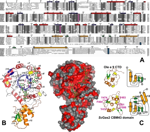
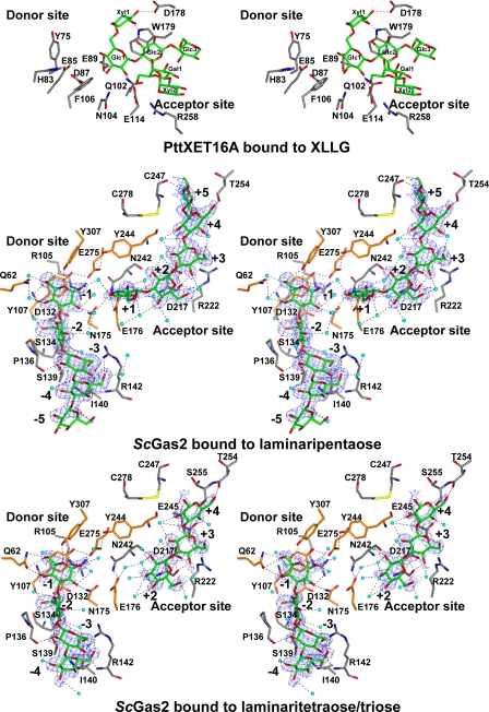
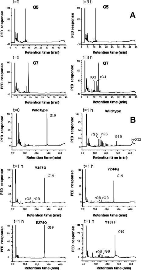
Yeast cell wall remodeling is controlled by the equilibrium between glycoside hydrolases, glycosyltransferases, and transglycosylases. Family 72 glycoside hydrolases (GH72) are ubiquitous in fungal organisms and are known to possess significant transglycosylase activity, producing elongated beta(1-3) glucan chains. However, the molecular mechanisms that control the balance between hydrolysis and transglycosylation in these enzymes are not understood. Here we present the first crystal structure of a glucan transglycosylase, Saccharomyces cerevisiae Gas2 (ScGas2), revealing a multidomain fold, with a (betaalpha)(8) catalytic core and a separate glucan binding domain with an elongated, conserved glucan binding groove. Structures of ScGas2 complexes with different beta-glucan substrate/product oligosaccharides provide "snapshots" of substrate binding and hydrolysis/transglycosylation giving the first insights into the mechanisms these enzymes employ to drive beta(1-3) glucan elongation. Together with mutagenesis and analysis of reaction products, the structures suggest a "base occlusion" mechanism through which these enzymes protect the covalent protein-enzyme intermediate from a water nucleophile, thus controlling the balance between hydrolysis and transglycosylation and driving the elongation of beta(1-3) glucan chains in the yeast cell wall.


Figures

FIGURE 1.
FIGURE 1.
Overall structure of ScGas2 and comparison with structurally related proteins. A, multiple sequence alignment of the GH72 family members ScGas2, ScGas1, CaPHR2, CaPHR1, AfGel3, AfGel1, and ScGas4. Secondary structure elements from the ScGas2 structure are shown, with α-helices in red and orange for the catalytic and cysteine-rich domains, respectively, and β-strands correspondingly in blue and green. Regions that are disordered in some or all of the ScGas2 structures are marked with a dashed line. Conserved catalytic glutamate residues are highlighted in pink boxing, and the (predicted) GPI-anchor attachment site is indicated in blue boxing. B, overall crystal structure of ScGas2 in complex with laminaripentaose. The Glu176 and Glu275 are shown with pink carbon atoms and labeled. The seven disulfide bridges are highlighted in yellow. Helix α13 was not built in the laminaripentaose complex structure and thus is absent from this figure. Ligand molecules are shown as sticks with green carbon atoms and the sugar-binding sites are labeled –5 to +5, following standard nomenclature. Other colors as in A. Also shown is a surface representation of the ScGas2, colored by sequence conservation (red (100% identity) to gray (<50% identity)). C, comparison of the CBM43 domains of ScGas2 (bottom; E176Q mutant) and Ole e 9 (top; PDB ID 2JON (39)). Disulfide bridge sulfur atoms are shown as yellow spheres. Secondary structure elements are colored as in B and labeled. Unique features of either structure are shown in lighter colors in the picture (left). The topology diagram was drawn with Topdraw (50). Surface-exposed aromatic amino acids of the CBM43 domain of ScGas2 (Phe404, Tyr417, Tyr474, Phe493, Tyr501, and Tyr506) are shown as sticks with pink carbon atoms.
FIGURE 2.
FIGURE 2.
Structures of ScGas2-laminarioligosaccharide complexes. Stereo view of the active site of ScGas2 in complex with laminaripentaose and the hydrolysis products of laminariheptaose (i.e. laminaritetraose + laminaritriose), and comparison with PttXET16A bound to XLLG. The active site oriented to facilitate identification of the donor (left) and acceptor (right) subsites. The amino acids placed in the donor site and acceptor sites are shown as sticks with gray carbons. The residues targeted by site-directed mutagenesis, Gln62, Tyr107, Asp132, Asn175, Glu176, Tyr244, Glu275, Tyr307, Phe404, and Tyr474, are shown with orange carbon atoms. XLLG, laminaripentaose, laminaritetraose, and laminaritriose are represented as stick models with green carbon atoms. Protein-ligand and water-ligand hydrogen bonds are shown as dotted black lines. Water molecules involved in hydrogen bonds with the ligands are shown as cyan spheres. For clarity purposes, protein-water hydrogen bonds are not shown. Unbiased (i.e. before inclusion of any ligand model) |Fo| – |Fc|, φcalc electron density maps are shown at 2.5 σ.
FIGURE 3.
FIGURE 3.
High pressure liquid chromatography analysis of β(1,3) glucanosyltransferase/hydrolysis products. A, comparison of wild type ScGas2 kinetics against laminaripentaose and laminariheptaose, identifying laminaritetraose and laminaritriose as the main two degradation products of hydrolyzed laminariheptaose. B, product analysis from the incubation of the recombinant wild type ScGas2 and the following single mutant enzymes, Y107F, Y244Q, E275Q, and Y307Q, with 4 mm reduced G19 samples taken at the indicated time points.

Similar articles

Cited by

References

    1. Latge, J. P. (2007) Mol. Microbiol. 66 279–290 - PubMed
    1. Fleet, G. H. (1991) in The Yeasts (Rose, A. H., ed) pp. 199–277, Academic Press, London
    1. Kollar, R., Petrakova, E., Ashwell, G., Robbins, P. W., and Cabib, E. (1995) J. Biol. Chem. 270 1170–1178 - PubMed
    1. Fontaine, T., Simenel, C., Dubreucq, G., Adam, O., Delepierre, M., Lemoine, J., Vorgias, C. E., Diaquin, M., and Latge, J. P. (2000) J. Biol. Chem. 275 27594–27607; Correction (2000) J. Biol. Chem. 275, 41528 - PubMed
    1. Adams, D. J. (2004) Microbiology 150 2029–2035 - PubMed

Publication types

MeSH terms

Substances