. 2008 Dec 30;105(52):20947-52.
doi: 10.1073/pnas.0804007106. Epub 2008 Dec 19.

Activity-independent and subunit-specific recruitment of functional AMPA receptors at neurexin/neuroligin contacts

Affiliations

Activity-independent and subunit-specific recruitment of functional AMPA receptors at neurexin/neuroligin contacts

Martin Heine et al. Proc Natl Acad Sci U S A. .

Abstract

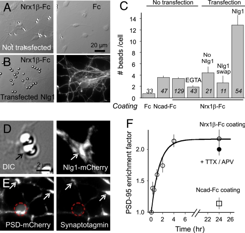
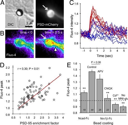
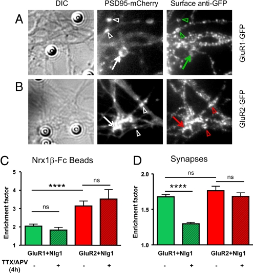
A combination of cell culture and animal studies has recently shown that adhesion between neurexins and neuroligins played important roles in synapse initiation, maturation, and function. Binding of neurexin-1beta to neuroligin-1 triggers the postsynaptic clustering of the scaffold postsynaptic density protein 95, but the composition and timing of accumulation of glutamate receptors at those nascent contacts remain unclear. Using glutamate iontophoresis and patch-clamp recordings, we identified functional AMPA receptors (AMPARs) and NMDA receptors at postsynaptic density protein 95 clusters induced by neurexin-1beta coated microspheres on primary hippocampal neurons. The recruitment of AMPARs occurred as early as 2 h after initial contact, and was not blocked by TTX/2-amino-5-phosphovaleric acid (APV) treatment. The differential recruitment of recombinant subunits GluR1 and GluR2, as well as the absence of rectification in voltage/current curves, further indicate that neurexin/neuroligin contacts primarily recruit GluR2-containing AMPARs. Finally, by using glutamate un-caging and calcium imaging, we show that AMPARs participate in calcium entry at neurexin-1beta induced post-synapses, most likely through the activation of voltage-gated calcium channels. Such rapid and activity-independent accumulation of functional AMPARs at neurexin-1beta-induced postsynapses points to a new role of AMPARs in synaptogenesis.


Conflict of interest statement

The authors declare no conflict of interest.

Figures

Fig. 1.
Fig. 1.
Binding of β-neurexin-coated beads and recruitment of postsynaptic proteins is neuroligin-specific. (A) Rat hippocampal neurons at 7 or 8 DIV were incubated with 4-μm microspheres coated with Nrx1β-Fc, N-cadherin-Fc (not shown), or human Fc for 1 h, then rinsed and fixed at given time intervals. Some cultures were co-transfected with PSD-95-GFP and WT Nlg1 (B) or an Nlg1 construct unable to interact with neurexin (Nlg1-SWAP). (C) Number of beads bound per cell in each condition. Data are presented as mean ± SEM and the number of beads examined is given in italics. (D) Example of recruitment of Nlg1-mCherry (arrows) at a Nrx1β-Fc bead. The enrichment factor (bead vs. neurite fluorescence levels) for Nlg1-mCherry was 1.71 ± 0.08 (n = 18 beads). (E) Example of recruitment of PSD-mCherry; position of the bead is represented by a dashed circle. Immuno-stained synaptotagmin puncta co-localize with spontaneous PSD-95 clusters (arrows), but not with PSD-95-mCherry accumulated at Nrx1β-Fc beads. Neuronal areas covered by synapses were 7.6% ± 1.0% (n = 13) outside beads and only 4.5% ± 1.3% (n = 48) at Nrx1β-Fc beads, quantitatively showing that Nrx1β-Fc beads do not recruit native synapses. (F) Time course of PSD-95-GFP accumulation at Nrx1β-Fc beads, the fit being a first-order exponential function.
Fig. 2.
Fig. 2.
Glutamate un-caging induces calcium transients at Nrx1β-Fc beads. Neurons co-transfected with Nlg1 and PSD-95-mCherry were incubated overnight with microspheres coated with Nrx1β-Fc. (A) Example of a Nrx1β-Fc bead having recruited PSD-95-mCherry. (B) Corresponding Fluo-4 signal increase upon photo-release of glutamate at time 0. (C) Representative examples of the normalized Fluo-4 fluorescence level over time for 20 beads. Traces in red correspond to beads showing significant PSD-95 recruitment, and those in blue correspond to beads with minimal PSD-95 recruitment. (D) Relationship between PSD-95 recruitment level and the peak in Fluo-4 signal elicited upon glutamate un-caging. (E) Effect of various treatments on the calcium response, presented as mean ± SEM, where the number of beads tested is indicated in italics. The dashed line represents no change in Fluo-4 level, and responses below this baseline indicate Fluo-4 photo-bleaching. Data were analyzed by one-way ANOVA and compared by Dunnett test to the control Nrx1β-Fc bead condition (**P < 0.01).
Fig. 3.
Fig. 3.
Characterization of glutamate receptor composition at Nrx1β-Fc beads by glutamate ionotophoresis and patch-clamp experiment. Neurons co-transfected with Nlg1 and PSD-95-GFP (or Homer1c-GFP) were left in contact with Nrx1β-Fc-coated microspheres for 2–4 h or 24 h, then subjected to whole-cell electrophysiology in the presence of TTX to prevent action potentials and 0 mM Mg to remove the NMDAR block. In some experiments, cells were treated with TTX and APV throughout the bead incubation (24 h). (A) Digital image correlation showing patch-clamp and iontophoresis pipettes, with the corresponding PSD-95-GFP fluorescence. Note that many beads bind to the transfected cell. (B) Magnified view of accumulation of PSD-95-GFP surrounding a Nrx1β-Fc bead and nearby synaptic clusters. (C) Examples of currents elicited upon glutamate application at Nrx1β-Fc beads (trace 1), endogenous PSD-95-GFP clusters (trace 2), or neurite region without PSD-95 accumulation (trace 3), with the membrane potential being clamped at −60 mV. (D) Effect of NMDAR and AMPAR antagonists (CNQX and APV, respectively) on the electrophysiological response of βNrx1β-Fc bead contacts at two contact durations. (E and F) By fitting the curves using a bi-exponential function with two separate time constants, the relative charge carried by AMPARs was computed for various conditions. (E) Synapses identified by PSD-95-GFP or Homer1c-GFP positive puncta. (F) Nrx1β-Fc beads displaying recruitment of PSD-95-GFP or Homer1c-GFP. Data were analyzed by one-way ANOVA and compared two-by-two by Tukey test (*, P < 0.05; ns, not significant).
Fig. 4.
Fig. 4.
Recruitment of recombinant AMPAR subunits at Nrx1β-Fc beads and synapses. Neurons were transfected with Nlg1 and PSD-95-mCherry plus either GluR1-GFP or GluR2-GFP, incubated for 4 h with Nrx1β-Fc beads with or without TTX/APV, then processed for live anti-GFP staining. Examples of recombinant GluR1 (A) or GluR2 (B) stainings at Nrx1β-Fc beads (arrows) and nearby endogenous synapses, as identified by PSD-95-mCherry clusters (arrowheads). Quantification of the enrichment factor, i.e., the fluorescence signal on a bead (C) or synapse (D) divided by the neurite level, for all conditions. The number of beads or clusters quantified is given in each column. Data were analyzed by one-way ANOVA and compared by Tukey test (***, P < 0.0002).
Fig. 5.
Fig. 5.
Comparison of postsynapses triggered by Nrx1β to endogenous synapses. (A) Nrx1β-Fc beads not exposed to chronic glutamate release induce the clustering of PSD-95, NMDAR, and GluR2-containing AMPAR, but exclude GluR2-lacking AMPAR receptors. (B) In contrast, synapses recruit GluR1-containing AMPAR in an activity-dependent manner.

References

    1. Missler M, Sudhof TC. Neurexins: three genes and 1001 products. Trends Genet. 1998;14:20–26. - PubMed
    1. Craig AM, Kang Y. Neurexin-neuroligin signaling in synapse development. Curr Opin Neurobiol. 2007;17:43–52. - PMC - PubMed
    1. Levinson JN, El-Husseini A. A crystal-clear interaction: relating neuroligin/neurexin complex structure to function at the synapse. Neuron. 2007;56:937–939. - PubMed
    1. Missler M, Fernandez-Chacon R, Sudhof TC. The making of neurexins. J Neurochem. 1998;71:1339–1347. - PubMed
    1. Ichtchenko K, et al. Neuroligin 1: a splice site-specific ligand for beta-neurexins. Cell. 1995;81:435–443. - PubMed

Publication types

MeSH terms