

LPS-induced autophagy is mediated by oxidative signaling in cardiomyocytes and is associated with cytoprotection
- PMID: 19098111
- PMCID: PMC2643899
- DOI: 10.1152/ajpheart.01051.2008
LPS-induced autophagy is mediated by oxidative signaling in cardiomyocytes and is associated with cytoprotection
Abstract
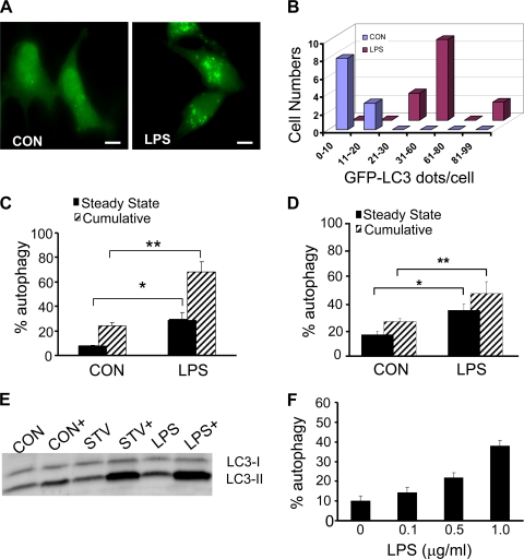
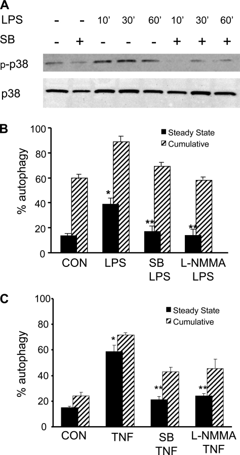
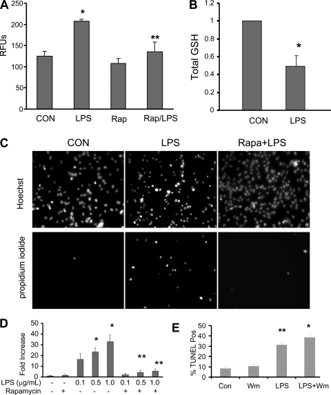
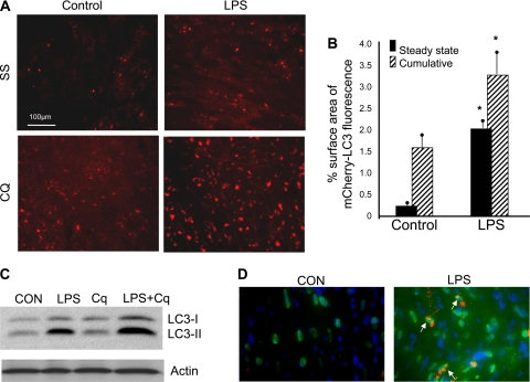
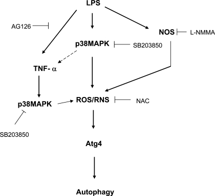
Bacterial endotoxin lipopolysaccharide (LPS) is responsible for the multiorgan dysfunction that characterizes septic shock and is causal in the myocardial depression that is a common feature of endotoxemia in patients. In this setting the myocardial dysfunction appears to be due, in part, to the production of proinflammatory cytokines. A line of evidence also indicates that LPS stimulates autophagy in cardiomyocytes. However, the signal transduction pathway leading to autophagy and its role in the heart are incompletely characterized. In this work, we wished to determine the effect of LPS on autophagy and the physiological significance of the autophagic response. Autophagy was monitored morphologically and biochemically in HL-1 cardiomyocytes, neonatal rat cardiomyocytes, and transgenic mouse hearts after the administration of bacterial LPS or TNF-alpha. We observed that autophagy was increased after exposure to LPS or TNF-alpha, which is induced by LPS. The inhibition of TNF-alpha production by AG126 significantly reduced the accumulation of autophagosomes both in cell culture and in vivo. The inhibition of p38 MAPK or nitric oxide synthase by pharmacological inhibitors also reduced autophagy. Nitric oxide or H(2)O(2) induced autophagy in cardiomyocytes, whereas N-acetyl-cysteine, a potent antioxidant, suppressed autophagy. LPS resulted in increased reactive oxygen species (ROS) production and decreased total glutathione. To test the hypothesis that autophagy might serve as a damage control mechanism to limit further ROS production, we induced autophagy with rapamycin before LPS exposure. The activation of autophagy by rapamycin suppressed LPS-mediated ROS production and protected cells against LPS toxicity. These findings support the notion that autophagy is a cytoprotective response to LPS-induced cardiomyocyte injury; additional studies are needed to determine the therapeutic implications.
Figures


References
-
- Brady NR, Hamacher-Brady A, Yuan H, Gottlieb RA. The autophagic response to nutrient deprivation in the HL-1 cardiac myocyte is modulated by Bcl-2 and sarco/endoplasmic reticulum calcium stores. FEBS J 274: 3184–3197, 2007. - PubMed
-
- Comstock KL, Krown KA, Page MT, Martin D, Ho P, Pedraza M, Castro EN, Nakajima N, Glembotski CC, Quintana PJ, Sabbadini RA. LPS-induced TNF-alpha release from and apoptosis in rat cardiomyocytes: obligatory role for CD14 in mediating the LPS response. J Mol Cell Cardiol 30: 2761–2775, 1998. - PubMed
-
- Cuervo AM Autophagy: in sickness and in health. Trends Cell Biol 14: 70–77, 2004. - PubMed
-
- Debnath J, Baehrecke EH, Kroemer G. Does autophagy contribute to cell death? Autophagy 1: 66–74, 2005. - PubMed
Publication types
MeSH terms
Substances
Grants and funding
LinkOut - more resources
Full Text Sources