

Glucosamine improves cardiac function following trauma-hemorrhage by increased protein O-GlcNAcylation and attenuation of NF-{kappa}B signaling
- PMID: 19098112
- PMCID: PMC2643896
- DOI: 10.1152/ajpheart.01025.2008
Glucosamine improves cardiac function following trauma-hemorrhage by increased protein O-GlcNAcylation and attenuation of NF-{kappa}B signaling
Abstract
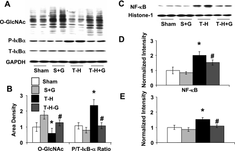
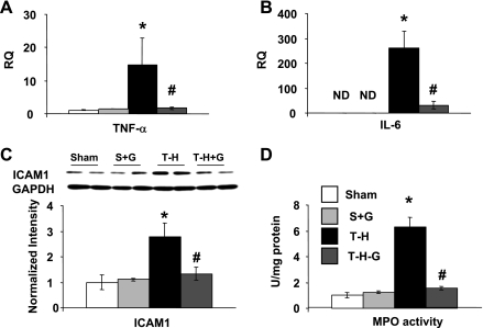
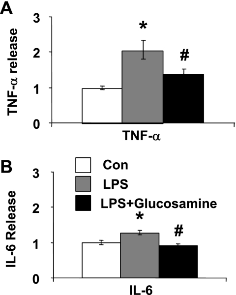
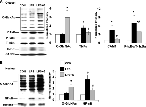
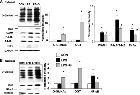
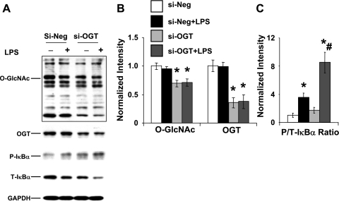
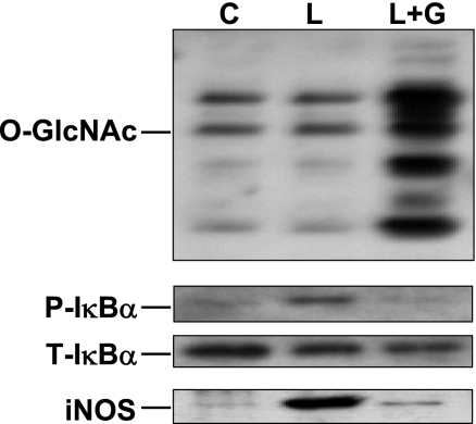
We have previously demonstrated that in a rat model of trauma-hemorrhage (T-H), glucosamine administration during resuscitation improved cardiac function, reduced circulating levels of inflammatory cytokines, and increased tissue levels of O-linked N-acetylglucosamine (O-GlcNAc) on proteins. The mechanism(s) by which glucosamine mediated its protective effect were not determined; therefore, the goal of this study was to test the hypothesis that glucosamine treatment attenuated the activation of the nuclear factor-kappaB (NF-kappaB) signaling pathway in the heart via an increase in protein O-GlcNAc levels. Fasted male rats were subjected to T-H by bleeding to a mean arterial blood pressure of 40 mmHg for 90 min followed by resuscitation. Glucosamine treatment during resuscitation significantly attenuated the T-H-induced increase in cardiac levels of TNF-alpha and IL-6 mRNA, IkappaB-alpha phosphorylation, NF-kappaB, NF-kappaB DNA binding activity, ICAM-1, and MPO activity. LPS (2 microg/ml) increased the levels of IkappaB-alpha phosphorylation, TNF-alpha, ICAM-1, and NF-kappaB in primary cultured cardiomyocytes, which was significantly attenuated by glucosamine treatment and overexpression of O-GlcNAc transferase; both interventions also significantly increased O-GlcNAc levels. In contrast, the transfection of neonatal rat ventricular myocytes with OGT small-interfering RNA decreased O-GlcNAc transferase and O-GlcNAc levels and enhanced the LPS-induced increase in IkappaB-alpha phosphorylation. Glucosamine treatment of macrophage cell line RAW 264.7 also increased O-GlcNAc levels and attenuated the LPS-induced activation of NF-kappaB. These results demonstrate that the modulation of O-GlcNAc levels alters the response of cardiomyocytes to the activation of the NF-kappaB pathway, which may contribute to the glucosamine-mediated improvement in cardiac function following hemorrhagic shock.
Figures
Similar articles
-
O-Linked β-N-Acetylglucosamine Modification of A20 Enhances the Inhibition of NF-κB (Nuclear Factor-κB) Activation and Elicits Vascular Protection After Acute Endoluminal Arterial Injury.Arterioscler Thromb Vasc Biol. 2018 Jun;38(6):1309-1320. doi: 10.1161/ATVBAHA.117.310468. Epub 2018 Apr 5. Arterioscler Thromb Vasc Biol. 2018. PMID: 29622561
-
[O-linked N-acetylglucosamine modification induced by lipopolysaccharide is involved in inflammatory signaling pathway in endothelial cells].Zhonghua Wei Zhong Bing Ji Jiu Yi Xue. 2023 Feb;35(2):164-169. doi: 10.3760/cma.j.cn121430-20220314-00242. Zhonghua Wei Zhong Bing Ji Jiu Yi Xue. 2023. PMID: 36916376 Chinese.
-
Glucosamine protects neonatal cardiomyocytes from ischemia-reperfusion injury via increased protein O-GlcNAc and increased mitochondrial Bcl-2.Am J Physiol Cell Physiol. 2008 Jun;294(6):C1509-20. doi: 10.1152/ajpcell.00456.2007. Epub 2008 Mar 26. Am J Physiol Cell Physiol. 2008. PMID: 18367586 Free PMC article.
-
Protein O-linked β-N-acetylglucosamine: a novel effector of cardiomyocyte metabolism and function.J Mol Cell Cardiol. 2012 Mar;52(3):538-49. doi: 10.1016/j.yjmcc.2011.08.009. Epub 2011 Aug 22. J Mol Cell Cardiol. 2012. PMID: 21878340 Free PMC article. Review.
-
O-GlcNAc signaling in the cardiovascular system.Circ Res. 2010 Jul 23;107(2):171-85. doi: 10.1161/CIRCRESAHA.110.224675. Circ Res. 2010. PMID: 20651294 Free PMC article. Review.
Cited by
-
Elevated O-GlcNAcylation promotes colonic inflammation and tumorigenesis by modulating NF-κB signaling.Oncotarget. 2015 May 20;6(14):12529-42. doi: 10.18632/oncotarget.3725. Oncotarget. 2015. PMID: 25915426 Free PMC article.
-
O-GlcNAcylation and Inflammation: A Vast Territory to Explore.Front Endocrinol (Lausanne). 2015 Jan 9;5:235. doi: 10.3389/fendo.2014.00235. eCollection 2014. Front Endocrinol (Lausanne). 2015. PMID: 25620956 Free PMC article. Review.
-
O-GlcNAcylation in immunity and inflammation: An intricate system (Review).Int J Mol Med. 2019 Aug;44(2):363-374. doi: 10.3892/ijmm.2019.4238. Epub 2019 Jun 11. Int J Mol Med. 2019. PMID: 31198979 Free PMC article. Review.
-
Cardiomyocyte Ogt is essential for postnatal viability.Am J Physiol Heart Circ Physiol. 2014 Jan 1;306(1):H142-53. doi: 10.1152/ajpheart.00438.2013. Epub 2013 Nov 1. Am J Physiol Heart Circ Physiol. 2014. PMID: 24186210 Free PMC article.
-
Stress-induced O-GlcNAcylation: an adaptive process of injured cells.Biochem Soc Trans. 2017 Feb 8;45(1):237-249. doi: 10.1042/BST20160153. Biochem Soc Trans. 2017. PMID: 28202678 Free PMC article. Review.
References
-
- Acosta JA, Yang JC, Winchell RJ, Simons RK, Fortlage DA, Hollingsworth-Fridlund P, Hoyt DB. Lethal injuries and time to death in a level I trauma center. J Am Coll Surg 186: 528–533, 1998. - PubMed
-
- Alam HB, Koustova E, Rhee P. Combat casualty care research: from bench to the battlefield. World J Surg 29, Suppl 1: S7–S11, 2005. - PubMed
-
- Angele MK, Schwacha MG, Ayala A, Chaudry IH. Effect of gender and sex hormones on immune responses following shock. Shock 14: 81–90, 2000. - PubMed
-
- Barringer ML, Thomason MH, Kilgo P, Spallone L. Improving outcomes in a regional trauma system: impact of a level III trauma center. Am J Surg 192: 685–689, 2006. - PubMed
Publication types
MeSH terms
Substances
Grants and funding
LinkOut - more resources
Full Text Sources
Molecular Biology Databases
Research Materials
Miscellaneous