. 2009 Mar;328(3):912-20.
doi: 10.1124/jpet.108.147959. Epub 2008 Dec 19.

Protein kinase Cbeta is a critical regulator of dopamine transporter trafficking and regulates the behavioral response to amphetamine in mice

Affiliations

Protein kinase Cbeta is a critical regulator of dopamine transporter trafficking and regulates the behavioral response to amphetamine in mice

Rong Chen et al. J Pharmacol Exp Ther. 2009 Mar.

Abstract

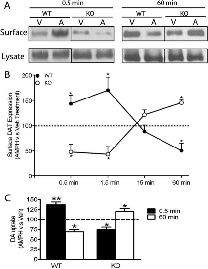
The dopamine transporter (DAT) is a key mediator of dopaminergic neurotransmission and a major target for amphetamine. We found previously that protein kinase C (PKC) beta regulates amphetamine-mediated dopamine efflux. Here, using PKCbeta wild-type (WT) and knockout (KO) mice, we report a novel role for PKCbeta in amphetamine-induced regulation of DAT trafficking and activity. PKCbeta KO mice have less striatal surface DAT, [3H]dopamine uptake, and amphetamine-stimulated dopamine efflux, yet higher novelty-induced locomotor activity than WT mice. Although a short exposure (< or =90 s) to amphetamine rapidly increases striatal surface DAT and [3H]dopamine uptake in WT mice, this treatment decreases surface DAT and [3H]dopamine uptake in KO mice. Increases in surface DAT and [3H]dopamine uptake are not evident in KO mice until a longer exposure (60 min) to amphetamine, by which time WT mice exhibit decreased surface DAT and dopamine uptake. The slowness of amphetamine-induced striatal DAT trafficking in PKCbeta KO mice was mimicked by the use of a specific PKCbeta inhibitor, LY379196, in WT mice. Furthermore, PKCbeta KO mice exhibit reduced locomotor responsiveness to amphetamine compared with WT, which could be explained by reduced surface DAT and delayed amphetamine-induced DAT trafficking in KO mice. Our results indicate that PKCbeta is crucial for proper trafficking of DAT to the surface and for functioning of DAT and amphetamine signaling, providing new insight into the role of PKCbeta as an important regulator of dopaminergic homeostasis.


Figures

Fig. 1.
Fig. 1.
Reduced striatal surface DAT expression in KO compared with WT mice. A, representative Western blot of the striatal surface and total DAT expression for WT and KO mice. Striatal synaptosomes were biotinylated, and bands were quantified to determine the surface and total DAT expression as described under Materials and Methods. KO mice showed less surface (biotinylated) DAT level than WT mice (Student's t test, p < 0.05, n = 6), whereas no significant genotype difference in the expression of the total DAT was observed. B, surface DAT was also compared between genotypes by determining binding of the DAT antagonist [3H]WIN 35,428 with striatal synaptosomes. Striatal synaptosomes from WT and KO mice were incubated with different concentrations of [3H]WIN35,428 for 1 h at 4 °C. Bmax was significantly lower for KO mice than for WT mice (Student's t test, p < 0.05, n = 5-6), whereas Kd values did not differ. Data are represented as mean ± S.E.M.
Fig. 2.
Fig. 2.
Reduced DAT activity in KO compared with WT mice. KO mice showed significantly less DA uptake (A) and AMPH-stimulated DA efflux (B) from striatal synaptosomes than WT mice. A, striatal synaptosomes from WT and KO mice were incubated with various concentrations of [3H]DA for 1 min at 37°C. The Vmax value for [3H]DA uptake was significantly lower in KO mice than in WT mice (Student's t test, p < 0.05, n = 6), whereas there was no difference in Km values. B, striatal synaptosomes from WT and KO mice were perfused (37°C) for 20 min with KRB at a speed of 450 μl/min before sample collections (2 min interval). Then, 1, 10, and 100 μM AMPH were introduced to the synaptosomes at fraction numbers 2, 8, and 14, respectively, and KRB was perfused for the rest of the fractions. A two-way ANOVA indicated a significant genotype effect [F(1,191) = 5.18, p < 0.01]. Post hoc Bonferroni analysis indicated that a significant reduction in AMPH induced DA efflux at fractions 3, 4, 9, 10, 15, and 16. *, p < 0.05; **, p < 0.01 WT versus KO. Data are represented as mean ± S.E.M.
Fig. 3.
Fig. 3.
Differential time-dependent DAT trafficking and DAT activity upon short- or long-term AMPH treatment between WT and KO mice. A, representative blots of surface (biotinylated fraction) and total DAT expression upon short- and long-term A (10 μM) or V treatment for WT and KO mice. B, surface DAT level was expressed as the ratio of biotinylated DAT versus the total DAT. The data shown in B were calculated as the percentage of surface DAT level upon the AMPH treatment (biotinylated DAT/lysate DAT) relative to the Veh treatment for WT and KO mice and graphically demonstrated as a time course. A two-way ANOVA (time × genotype) revealed a significant interaction effect of genotype and time [F(3,42) = 10.90, p < 0.01]. Upon short-term AMPH exposure (0.5 and 1.5 min), there was a significant increase in surface DAT expression in striatal synaptosomes for WT mice (n = 8) and a significant decrease for KO mice (n = 5) compared with their control (Veh-treated) groups (AMPH versus Veh, paired Student's t test; *, p < 0.05). Upon long-term AMPH exposure (60 min), there was a significant decrease in surface DAT expression for WT mice (n = 5) and a significant increase for KO mice (n = 3) (AMPH versus Veh, paired Student's t test; *, p < 0.05). C, striatal synaptosomes were pretreated with AMPH or Veh for 0.5 or 60 min. Then, synaptosomes were washed extensively to remove residual AMPH before measurement of [3H]DA uptake. Data were expressed as the percentage of the [3H]DA uptake into synaptosomes pretreated with AMPH relative to Veh. Upon short-term AMPH exposure (0.5 min), there was a significant increase in [3H]DA uptake into synaptosomes from WT mice compared with their Veh-treated control groups (paired Student's t test, p < 0.01, n = 4) and a significant decrease for KO mice (paired Student's t test, p < 0.05, n = 4). Upon long-term AMPH exposure (60 min), there was a significant decrease in [3H]DA uptake into synaptosomes from WT mice (n = 6) and a significant increase for KO mice (n = 5) compared with control synaptosomes. *, p < 0.05; **, p < 0.01. Data are represented as mean ± S.E.M.
Fig. 4.
Fig. 4.
Effect of PKCβ inhibition on basal and AMPH-induced DAT trafficking in WT mice. Striatal synaptosomes from WT mice were pretreated with either the PKCβ-specific inhibitor LY (100 nM) or V for 1 h at 37°C. Surface DAT expression was determined by biotinylation and quantified as described under Materials and Methods. Data were expressed as percentage of DAT surface expression (biotinylated DAT/lysate DAT) relative to control. A, incubation with 100 nM LY for 1 h at 37°C significantly reduced basal surface DAT expression compared with Veh treatment (LY versus V, paired Student's t test, p < 0.05, n = 9). B, striatal synaptosomes from WT mice were preincubated with 100 nM LY379196, then challenged with 10 μM A or V for either 0.5 or 60 min. Short-term (0.5 min) AMPH exposure resulted in a significant reduction in surface DAT expression compared with Veh exposure (LY + V versus LY + 30-sec AMPH, paired Student's t test, p < 0.05, n = 4), mimicking short-term AMPH-induced DAT trafficking in KO mice. Conversely, long-term AMPH treatment (60 min) increased surface DAT expression compared with Veh controls (LY + V versus LY + 60-min AMPH, paired Student's t test, p < 0.01, n = 6), a similar pattern to KO mice. Data are represented as mean ± S.E.M.
Fig. 5.
Fig. 5.
Increased novelty-induced locomotor activity in KO mice compared with WT mice. WT and KO mice (n = 6) were placed in a novel environment, and locomotor activity was recorded over 2 h. A two-way ANOVA (genotype × time) revealed a significant main effect of genotype [F(1,207) = 20.27, p < 0.01]. KO mice showed significantly higher locomotor activity than WT mice. Data are represented as mean ± S.E.M.
Fig. 6.
Fig. 6.
Decreased responsiveness to AMPH in KO mice compared with WT mice. A, time course of locomotor activity induced by 3 mg/kg AMPH or Sal in WT and KO mice. Mice were given an injection of Sal to reduce injection stress 2 h before either AMPH (3 mg/kg) or Sal, and locomotor activity was recorded for 4 h as described under Materials and Methods. A two-way ANOVA (genotype × time) comparing AMPH-treated WT and KO mice revealed a significant main effect of genotype [F(1,1076) = 28.31, p < 0.0001], time [F(75,1076) = 17.45, p < 0.0001], and a significant interaction effect of genotype and time [F(75,1076) = 1.602, p < 0.001]. KO mice exhibited significantly lower locomotor activity in response to 3 mg/kg AMPH than WT mice (n = 8-9). Bar, significant values determined by post hoc Bonferroni analysis. *, p < 0.05 for all values except for 50 min (**, p < 0.01). B, mice were given an injection of Sal 2 h before the test injection of Sal or AMPH at doses of 1, 2, 3, 5, or 7 mg/kg. Locomotor activity was summed over 80 min. A two-way ANOVA (genotype × dose) revealed a significant main effect of AMPH dose [F(5, 87) = 61.49, p < 0.01] and a significant interaction effect of genotype and dose [F(5, 87) = 4.79, p < 0.05]. Post hoc Bonferroni analysis indicated that AMPH significantly stimulated locomotor activity in WT mice at doses of 1, 2, 3, 5, and 7 mg/kg compared with their Sal injection (n = 6-12), whereas KO mice showed stimulation only at doses of 3, 5, and 7 mg/kg AMPH (n = 6-13). In addition, the Sal-treated KO mice consistently exhibited higher locomotor activity than Sal-treated WT mice (Student's t test, p < 0.05, n = 11-13). *, p < 0.05; **, p < 0.01, AMPH versus Sal within each genotype; #, p < 0.05, WT versus KO within each dose of AMPH.

References

    1. Blobe GC, Stribling DS, Fabbro D, Stabel S, and Hannun YA (1996) Protein kinase C beta II specifically binds to and is activated by F-actin. J Biol Chem 271 15823-15830. - PubMed
    1. Bolan EA, Kivell B, Jaligam V, Oz M, Jayanthi LD, Han Y, Sen N, Urizar E, Gomes I, Devi LA, et al. (2007) D2 receptors regulate dopamine transporter function via an extracellular signal-regulated kinases 1 and 2-dependent and phosphoinositide 3 kinase-independent mechanism. Mol Pharmacol 71 1222-1232. - PubMed
    1. Boudanova E, Navaroli DM, and Melikian HE (2008) Amphetamine-induced decreases in dopamine transporter surface expression are protein kinase C-independent. Neuropharmacology 54 605-612. - PMC - PubMed
    1. Browman KE, Kantor L, Richardson S, Badiani A, Robinson TE, and Gnegy ME (1998) Injection of the protein kinase C inhibitor Ro31-8220 into the nucleus accumbens attenuates the acute response to amphetamine: tissue and behavioral studies. Brain Res 814 112-119. - PubMed
    1. Cervinski MA, Foster JD, and Vaughan RA (2005) Psychoactive substrates stimulate dopamine transporter phosphorylation and down-regulation by cocaine-sensitive and protein kinase C-dependent mechanisms. J Biol Chem 280 40442-40449. - PubMed

Publication types

MeSH terms