. 2009 May;50(5):966-76.
doi: 10.1194/jlr.M800632-JLR200. Epub 2008 Dec 20.

Nuclear export of the rate-limiting enzyme in phosphatidylcholine synthesis is mediated by its membrane binding domain

Affiliations

Nuclear export of the rate-limiting enzyme in phosphatidylcholine synthesis is mediated by its membrane binding domain

Karsten Gehrig et al. J Lipid Res. 2009 May.

Abstract

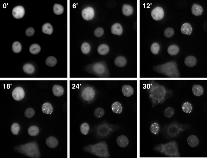
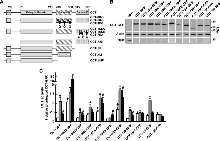
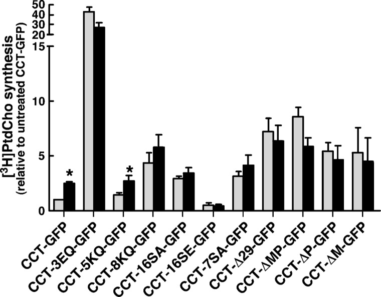
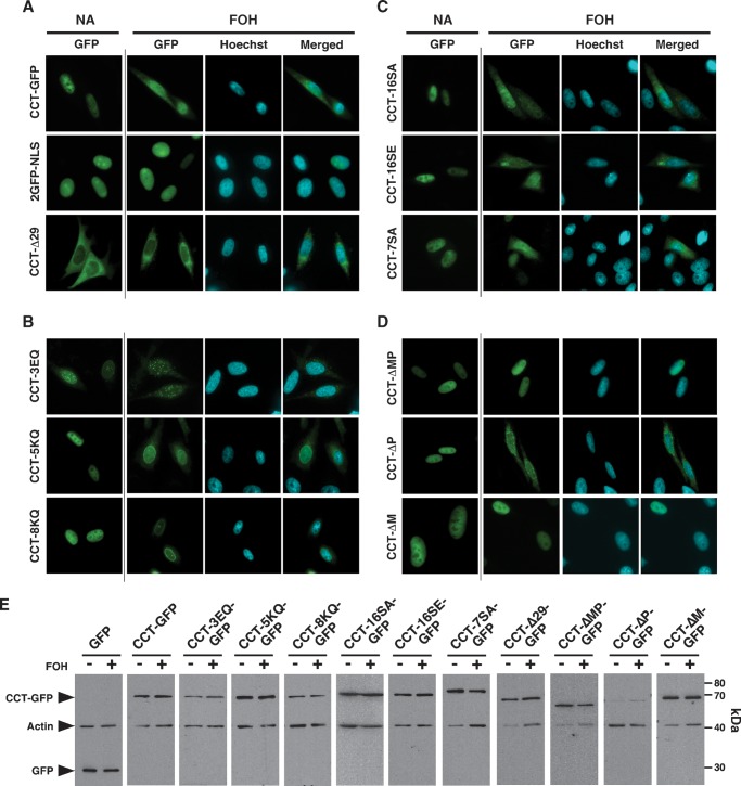
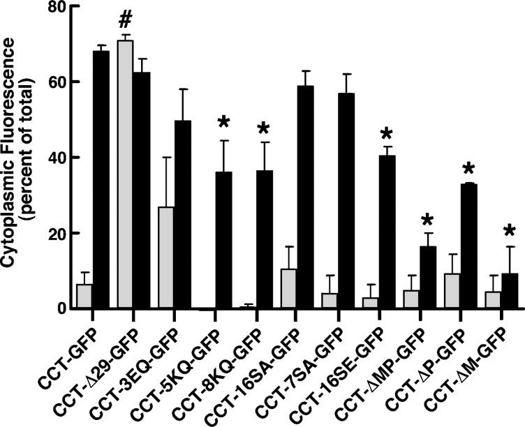
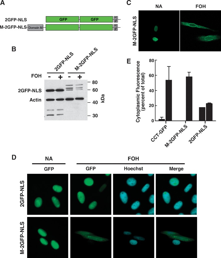
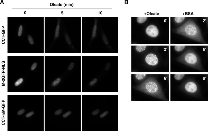
CTP:phosphocholine cytidylyltransferase alpha (CCTalpha), the rate-limiting enzyme in the CDP-choline pathway for phosphatidylcholine (PtdCho) synthesis, is activated by translocation to nuclear membranes. However, CCTalpha is cytoplasmic in cells with increased capacity for PtdCho synthesis and following acute activation, suggesting that nuclear export is linked to activation. The objective of this study was to identify which CCTalpha domains were involved in nuclear export in response to the lipid activators farnesol (FOH) and oleate. Imaging of CCT-green fluorescent protein (GFP) mutants expressed in CCTalpha-deficient CHO58 cells showed that FOH-mediated translocation to nuclear membranes and export to the cytoplasm required the membrane binding amphipathic helix (domain M). Nuclear export was reduced by a mutation that mimics constitutive phosphorylation of the CCT phosphorylation (P) domain. However, domain M alone was sufficient to promote translocation to the nuclear envelope and export of a nuclear-localized GFP construct in FOH- or oleate-treated CHO58 cells. In the context of acute activation with lipid mediators, nuclear export of CCT-GFP mutants correlated with in vitro activity but not PtdCho synthesis. This study describes a nuclear export pathway that is dependent on membrane interaction of an amphipathic helix, thus linking lipid-dependent activation to the nuclear/cytoplasmic distribution of CCTalpha.


Figures

Fig. 1.
Fig. 1.
Nuclear export of cytidine triphosphate:phosphocholine cytidylyltransferase-green fluorescent protein (CCT-GFP) in live cells. CHO58 cells transiently expressing CCT-GFP were mounted on a 40°C heated microscope stage. After equilibration for 15–20 min, cells received 60 μM farnesol (FOH), and images were captured at 30 s intervals (150 ms exposures) for 30 min as described in Materials and Methods. The Quicktime movie from which these individual images were extracted is shown in supplementary Fig. I.
Fig. 2.
Fig. 2.
In vitro activity and expression of CCT-GFP mutants. A: Domain structure of wild-type and CCTα mutants. GFP was fused to the C terminus of all constructs (not shown). B: CHO58 cells were transfected with CCT-GFP or the indicated mutants at 37°C for 12–14 h, shifted to 40°C or 42°C for 2 h and harvested, and 16,000 g supernatants were prepared and immunoblotted for GFP and actin. C: Enzyme activity was assayed in cell supernatants in the absence of lipid activators (open bars), with phosphatidylcholine (PtdCho)-oleate vesicles (1:1, mol/mol) (grey bars), or with PtdCho-FOH vesicle (3:2, mol/mol) (black bars) as described in Materials and Methods. Background CCT activity in supernatants from cells transfected with pEGFP was subtracted. Activity of wild-type and CCT mutants was normalized to expression of individual CCT-GFP proteins (measured by immunoblotting) and then expressed relative to activity under unstimulated conditions. CCT activity averaged 1,288, 1,091, and 1,217 dpm/assay in the supernatants of pEGFP-transfected cells without addition, plus oleate, or plus FOH, respectively. Activity averaged 1,653, 7,138, and 4,124 dpm/assay in supernatants from CCT-GFP-expressing cells with no addition, plus oleate, or plus FOH, respectively. Results are the mean and SEM of three separate experiments. *P < 0.05 compared with corresponding unstimulated activity; #P < 0.05 compared with corresponding unstimulated activity; ♦P < 0.05 compared with unstimulated CCT-GFP.
Fig. 3.
Fig. 3.
PtdCho synthesis in CHO58 cells expressing CCT-GFP mutants. CHO58 cells were transfected with CCT-GFP or the indicated mutants as described in the legend to Fig. 2. After 2 h at 42°C, cells were incubated with choline-free DMEM containing [3H]choline (2 μCi/ml) for 1 h, followed by treatment without (grey bars) or with (black bars) 300 μM oleate/complexed with BSA for 1 h. [3H]PtdCho was measured and normalized to expression of individual CCT-GFP proteins by immunoblotting of whole-cell lysates, and expressed relative to synthesis in unstimulated CHO58 cells transfected with CCT-GFP. Background [3H]PtdCho synthesis in cells transfected with pEGFP was subtracted. Total PtdCho synthesis was 1,045 and 910 dpm/dish in control and oleate-treated pEGFP-transfected cells, respectively. Total PtdCho synthesis was 13,655 and 36,550 dpm/dish in control and oleate-treated CCT-GFP-expressing cells, respectively. *P < 0.05 compared with corresponding unstimulated control. Results are the mean and SEM of three separate experiments.
Fig. 4.
Fig. 4.
Localization of wild-type and CCT-GFP mutants in FOH-treated CHO58 cells. Vectors encoding CCT-GFP or the indicated mutant proteins were expressed in CHO58 cells for 14 h, and shifted to 40°C for 1 h prior to stimulation with 60 μM FOH. After 45 min, cells were fixed and GFP and the nucleus (Hoechst staining) were visualized. A: CCT-GFP and nuclear 2GFP-nuclear localization signal (2GFP-NLS) and cytoplasmic (CCT-Δ29-GFP)-localized controls. B: Domain M point mutants (CCT-8KQ-GFP, -5KQ-GFP, and -3EQ-GFP). C: Phosphorylation mutants (CCT-16SA-GFP, -16SE-GFP, and -7SA-GFP). D: Domain M and P truncation mutants (CCT-ΔM-GFP, -ΔP-GFP, and -ΔMP-GFP). E: Total cell lysates prepared from CHO58 cells, transfected and treated with FOH as described above, were resolved by SDS-PAGE and immunoblotted for GFP and actin.
Fig. 5.
Fig. 5.
Quantification of nuclear export reveals a role for domain M. Vectors encoding wild-type and mutant CCT-GFP were transfected in CHO58 cells for 14 h and imaged following FOH treatment as described in the legend to Fig. 4. The percent distribution of GFP fluorescence in the cytoplasm of at least 20 control (grey bars) and FOH-treated (black bars) cells was quantified for each GFP fusion protein as described in Materials and Methods. *P < 0.05 compared with FOH treated CCT-GFP; #P < 0.05 compared with untreated CCT-GFP cells. Results are the mean and SEM of three separate experiments.
Fig. 6.
Fig. 6.
Domain M is necessary and sufficient for nuclear membrane translocation and export. A: Tandem repeats of GFP were targeted to the nucleus by attachment of the SV40 NLS (2GFP-NLS). Domain M was attached to the N terminus to make the M-2GFP-NLS construct. B: Total lysates of CHO58 cells transiently expressing 2GFP-NLS or M-2GFP-NLS for 14 h and treated with or without 60 μM FOH for 45 min were immunoblotted for GFP and actin. C: Confocal imaging of M-2GFP-NLS in CHO58 cells treated with and without 60 μM FOH for 45 min. D: Wide-field fluorescence micrographs of M-2GFP-NLS or 2GFP-NLS exposed to 60 μM FOH for 45 min. The nucleus was visualized by Hoechst staining. E: Quantification of the cytoplasmic distribution of CCT-GFP, 2GFP-NLS, and M-2GFP-NLS fluorescence in control (grey bars) and FOH-treated (black bars) cells shown in D. Results are the mean and SEM of three separate experiments that involved analysis of at least 20 cells each.
Fig. 7.
Fig. 7.
Oleate promotes domain M-dependent export of CCT-GFP. A: CHO58 cells expressing CCT-GFP, CCT-ΔM-GFP, or M-2GFP-NLS were prepared for live-cell imaging as described in Materials and Methods. Cells were mounted on a 37°C heated stage and treated with an oleate-BSA complex (300 μM oleate), and images were captured (750 ms exposures) at the indicated times. B: CHO58 cells expressing CCT-GFP were treated with oleate (320 μM complexed with BSA) at 40°C, and images were captured (150 ms exposures) at 30 s intervals for 6 min. Cells that displayed significant nuclear export of CCT-GFP were identified, and medium was replaced with prewarmed (40°C) oleate-free F-12 medium containing 0.2% BSA. Images were captured for a further 9 min. The Quicktime movie from which these images were taken is shown in supplementary Fig. III.

Similar articles

Cited by

References

    1. Vance J. E., and D. E. Vance. 2005. Metabolic insights into phospholipid function using gene-targeted mice. J. Biol. Chem. 280 10877–10880. - PubMed
    1. Cui Z., and M. Houweling. 2002. Phosphatidylcholine and cell death. Biochim. Biophys. Acta. 1585 87–96. - PubMed
    1. Vance D. E., Z. Li, and R. L. Jacobs. 2007. Hepatic phosphatidylethanolamine N-methyltransferase, unexpected roles in animal biochemistry and physiology. J. Biol. Chem. 282 33237–33241. - PubMed
    1. Lykidis A., and S. Jackowski. 2001. Regulation of mammalian cell membrane biosynthesis. Prog. Nucleic Acid Res. Mol. Biol. 65 361–393. - PubMed
    1. Karim M., P. Jackson, and S. Jackowski. 2003. Gene structure, expression and identification of a new CTP:phosphocholine cytidylyltransferase beta isoform. Biochim. Biophys. Acta. 1633 1–12. - PubMed

Publication types

MeSH terms

Substances

LinkOut - more resources