. 2009 Mar 13;284(11):7214-22.
doi: 10.1074/jbc.M805890200. Epub 2008 Dec 20.

Cyst formation in kidney via B-Raf signaling in the PKD2 transgenic mice

Affiliations

Cyst formation in kidney via B-Raf signaling in the PKD2 transgenic mice

Eun Young Park et al. J Biol Chem. .

Abstract

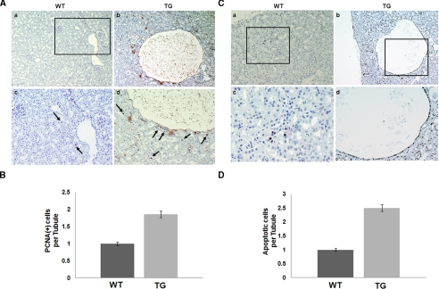
The pathogenic mechanisms of human autosomal dominant polycystic kidney disease (ADPKD) have been well known to include the mutational inactivation of PKD2. Although haploinsufficiency and loss of heterozygosity at the Pkd2 locus can cause cyst formation in mice, polycystin-2 is frequently expressed in the renal cyst of human ADPKD, raising the possibility that deregulated activation of PKD2 may be associated with the cystogenesis of human ADPKD. To determine whether increased PKD2 expression is physiologically pathogenic, we generated PKD2-overexpressing transgenic mice. These mice developed typical renal cysts and an increase of proliferation and apoptosis, which are reflective of the human ADPKD phenotype. These manifestations were first observed at six months, and progressed with age. In addition, we found that ERK activation was induced by PKD2 overexpression via B-Raf signaling, providing a possible molecular mechanism of cystogenesis. In PKD2 transgenic mice, B-Raf/MEK/ERK sequential signaling was up-regulated. Additionally, the transgenic human polycystin-2 partially rescues the lethality of Pkd2 knock-out mice and therefore demonstrates that the transgene generated a functional product. Functional strengthening or deregulated activation of PKD2 may be a direct cause of ADPKD. The present study provides evidence for an in vivo role of overexpressed PKD2 in cyst formation. This transgenic mouse model should provide new insights into the pathogenic mechanism of human ADPKD.


Figures

FIGURE 1.
FIGURE 1.
PKD2 transgenic mice. A, schematic map of the human cytomegalovirus enhancer and the human PKD2 cDNA construct. Restriction enzyme sites and probes used for Southern hybridization are indicated. B, copy number in PKD2 transgenic mice. Genomic DNA was subjected to real-time PCR and signals were normalized using Gapdh as an internal control. Endogenous signals were ruled out from the average of triplicate experiments, and transgene copy number per genome was expressed from triplicate experiments. Error bar, standard deviation. C, semiquantitative reverse transcriptase-PCR analysis was performed using a human PKD2-specific primer pair. The expression of the PKD2 transgene was determined in kidneys from four-independent transgenic mouse lines. D, Western blot analysis of polycystin-2 in kidneys of PKD2 transgenic mice. Polycystin-2 expression was evaluated in four-independent transgenic mouse lines. The polycystin-2-specific antibody could detect both human and mouse orthologues. WT, wild-type mouse; C, D, E, and F, transgenic mice lines.
FIGURE 2.
FIGURE 2.
Cystic phenotype in kidneys of PKD2 transgenic mice. H&E staining of PKD2 transgenic mouse kidney. A, the enlarged kidney filled with fluid from an 18-month-old PKD2 transgenic mouse. B and C, cysts were generated in variable regions of the kidney. B, a kidney section of an affected 18-month-old PKD2 transgenic mouse shows variable cyst size. Cysts arose in the cortex (C*) and medulla (M*). C, the morphological change of glomeruli (G*) in an 18-month-old PKD2 transgenic mouse. D-F, morphological changes of cyst-lining epithelial cells in the PKD2 transgenic mouse kidney during the development of renal cysts. D, at the early stage of cyst formation, tubules lined by cuboidal epithelium. E, cyst at the intermediate stage of formation, where epithelial cells are transformed into flattened cells (black arrowhead). F, magnification of the intermediate stage cyst. G and H, at the later stage of cyst formation. G, the cuboidal epithelium is completely transformed into flat, single-layered lining cells. H, magnification of the late stage cyst. C*, cortical cyst; M*, cyst in medullar region; G*, glomerular alteration; scale bar, 50 μm.
FIGURE 3.
FIGURE 3.
Sustained expression of polycystin-2 in the renal cysts of PKD2 transgenic mice. Immunohistochemical staining of polycystin-2 in kidneys of wild-type (WT)(A and B) and PKD2 transgenic (TG) mice (C and D). Boxed regions (A and C) were magnified (B and D). Polycystin-2 expression was detected in basolateral regions of distal tubules in wild-type (WT) kidney (A and B). Polycystin-2 was strongly expressed in the large cyst of the transgenic mouse (C and D). Scale bar, 50 μm.
FIGURE 4.
FIGURE 4.
Variable origins of cysts in PKD2 transgenic mice. The origins of the cystic lesions in PKD2 transgenic mice were immunohistochemically defined. A and B, expression of L. tetragonolobus (LTA, the proximal tubule marker) was detected in one of the cysts and surrounding dilated tubules (A). C and D, the cyst that originated from collecting tubules was identified by detecting the expression of D. biflorus (collecting tubule marker). Note that the cysts on serial sections were alternatively stained by L. tetragonolobus and D. biflorus (A and C). E and F, the large cyst positively stained with Tamm-Horsfall protein (THP)-specific antibody (distal tubule marker). Boxed regions (E) were magnified (F). *, cyst; scale bar, 50 μm.
FIGURE 5.
FIGURE 5.
Progressive deterioration of cystic phenotypes with aging. The mean sizes of cysts are shown for all PKD2 transgenic mouse lines according to age. Note that more cysts exceeding 200 μm were found in aged transgenic mice (18 months) than in young transgenic mice (6 months). Cystic tubules larger than 50 μm in diameter were counted, and the morphology of epithelial cells lining the cysts was evaluated (*, p < 0.01).
FIGURE 6.
FIGURE 6.
Increase of tubular cell proliferation and apoptosis in the cystic kidneys of PKD2 transgenic mice. PCNA-positive cells in the tubules of the cortex and medulla were significantly increased in PKD2 transgenic mice. Specifically, considerable numbers of PCNA-positive cells were also cyst-lining cells, and were observed in surrounding regions. A, a and c, kidney of a wild-type mouse. b and d, kidney of a PKD2 transgenic mouse. Boxed regions are magnified panels c and d. Note that flattened cells in the cyst were stained with PCNA. B, quantification of PCNA positive cells in wild-type (WT) and PKD2-transgenic (TG) kidneys, showing that the number of PCNA-positive cells per tubule was significantly increased in the PKD2 transgenic kidney compared with that of wild-type mice. The apoptotic index was evaluated by TUNEL assay. The number of apoptotic cells in the cysts and tubules around cysts were significantly increased in the PKD2 transgenic mouse compared with that of wild-type mice. C, a and c, kidney of a wild-type mouse. b and d, kidney of a PKD2 transgenic mouse. Boxed regions were magnified (c and d). Note that, similar to cell proliferation, apoptotic cells were enriched in cyst-lining cuboidal cells. D, quantification of TUNEL positive cells.
FIGURE 7.
FIGURE 7.
Activation of B-Raf/MEK/ERK signaling in the cystic kidneys of PKD2 transgenic mice. A, B-Raf/MEK/ERK sequential signaling was induced in PKD2 transgenic (TG) mice kidneys. The levels of phosphorylated and total B-Raf/MEK/ERK were examined by Western blot analysis. β-Actin was used as a loading control. PC2, polycystin-2. B, the graph shows quantification data of A. C, the statue of Akt phosphorylation was altered in PKD2 transgenic mice kidneys. D, the levels of phosphorylated ERK was reduced by transfection of B-Raf small interfering RNA and these levels were similar to the results of the MEK inhibitor treatment (PD98059). E, ERK phosphorylation level was also elevated in PKD2 transgenic mice but the phosphorylation level of p38 MAPK and JNK1/2 were unchanged in PKD2 transgenic mice. WT, wild type.
FIGURE 8.
FIGURE 8.
Abnormal activation of ERK signaling in the cystic kidneys of PKD2 transgenic mice. A, the statue of ERK phosphorylation was altered in PKD2-transgenic MEFs and kidneys. The levels of phosphorylated and total ERK were examined by Western blot analysis. β-Actin was used as a loading control. MEFs were derived from each PKD2 transgenic line (F embryo and Pkd2 deficient embryo). B-D, immunohistochemical analysis of phosphorylated ERK1/2 of wild-type (WT) kidneys in the control mouse (B) and cystic kidneys (C and D) in the PKD2 transgenic mouse. Boxed region in C was magnified in D. Note that phosphorylated ERK1/2, localized in nuclei, was detected in cyst-lining epithelium and tubules around the cysts. Scale bar, 50 μm. E, ERK1/2 was activated by exogenous PKD2 expression in wild-type MEFs.

Similar articles

Cited by

References

    1. Gabow, P. A. (1993) N. Engl. J. Med. 329 332-342 - PubMed
    1. Grantham, J. J. (1996) Am. J. Kidney Dis. 28 788-803 - PubMed
    1. Carone, F. A., Bacallao, R., and Kanwar, Y. S. (1996) The etiology, pathogenesis, and treatment of autosomal dominant polycystic kidney disease: recent advances, pp. 111-124, Oxford University Press, Oxford, UK
    1. Peters, D. J., and Sandkuijl, L. A. (1992) Contrib. Nephrol. 97 128-139 - PubMed
    1. Roscoe, J. M., Brissenden, J. E., Williams, E. A., Chery, A. L., and Silverman, M. (1993) Kidney Int. 44 1101-1108 - PubMed

Publication types

MeSH terms

Substances