Disparity tuning of binocular facilitation and suppression after normal versus abnormal visual development
- PMID: 19098323
- PMCID: PMC3637964
- DOI: 10.1167/iovs.08-2281
Disparity tuning of binocular facilitation and suppression after normal versus abnormal visual development
Abstract
Purpose: To study the pattern of facilitatory and suppressive binocular interactions in stereodeficient patients with strabismus and in healthy controls.
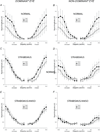
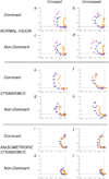
Methods: Visual evoked potentials were recorded in response to a Vernier onset/offset pattern presented to one eye, either monocularly or paired dichoptically with a straight vertical square-wave grating, which, when fused with the target in the other eye, gave rise to a percept of a series of bands appearing in depth from an otherwise uniform plane or with a grating that contained offsets that produced a standing disparity and the appearance of a constantly segmented image, portions of which moved in depth.
Results: Participants with normal stereopsis showed facilitative and suppressive binocular interactions that depended on which dichoptic target was presented. Patients with longstanding, constant strabismus lacked normal facilitative binocular interactions. The response to a normally facilitative stimulus was reduced below the monocular level when it was presented to the dominant eye of patients without anisometropia, consistent with classical strabismic suppression of the nondominant eye. The dominant eye of strabismic patients without anisometropia retained suppressive input from crossed but not uncrossed disparity stimuli presented to the nondominant eye.
Conclusions: Abnormal disparity processing can be detected with the dichoptic VEP method we describe. Our results suggest that suppression in stereoblind, nonamblyopic observers is determined by a binocular mechanism responsive to disparity. In some cases, the sign of the disparity is important, and this suggests a mechanism that can explain diplopia in patients made exotropic after surgery for esotropia.
Figures


Similar articles
-
A VEP measure of the binocular fusion of horizontal and vertical disparities.Invest Ophthalmol Vis Sci. 2005 May;46(5):1786-90. doi: 10.1167/iovs.04-0954. Invest Ophthalmol Vis Sci. 2005. PMID: 15851583
-
Excitatory Contribution to Binocular Interactions in Human Visual Cortex Is Reduced in Strabismic Amblyopia.J Neurosci. 2021 Oct 13;41(41):8632-8643. doi: 10.1523/JNEUROSCI.0268-21.2021. Epub 2021 Aug 25. J Neurosci. 2021. PMID: 34433631 Free PMC article.
-
Development of rivalry and dichoptic masking in human infants.Invest Ophthalmol Vis Sci. 1999 Dec;40(13):3324-33. Invest Ophthalmol Vis Sci. 1999. PMID: 10586959
-
Development of human binocular vision: An electrophysiological perspective.Vision Res. 2025 Jun;231:108593. doi: 10.1016/j.visres.2025.108593. Epub 2025 Apr 15. Vision Res. 2025. PMID: 40239434 Review.
-
Psychophysics of suppression.Eye (Lond). 1996;10 ( Pt 2):270-3. doi: 10.1038/eye.1996.57. Eye (Lond). 1996. PMID: 8776459 Review.
Cited by
-
Comparison of binocular visual function among patients with different types of anisometropia.Adv Ophthalmol Pract Res. 2025 Jun 18;5(3):182-188. doi: 10.1016/j.aopr.2025.04.005. eCollection 2025 Aug-Sep. Adv Ophthalmol Pract Res. 2025. PMID: 40678193 Free PMC article.
-
Altered spontaneous brain activity in patients with strabismic amblyopia: A resting-state fMRI study using regional homogeneity analysis.Exp Ther Med. 2019 Nov;18(5):3877-3884. doi: 10.3892/etm.2019.8038. Epub 2019 Sep 23. Exp Ther Med. 2019. PMID: 31616514 Free PMC article.
-
Alternating fixation and saccade behavior in nonhuman primates with alternating occlusion-induced exotropia.Invest Ophthalmol Vis Sci. 2009 Aug;50(8):3703-10. doi: 10.1167/iovs.08-2772. Epub 2009 Mar 11. Invest Ophthalmol Vis Sci. 2009. PMID: 19279316 Free PMC article.
References
-
- Tyler CW. Stereoscopic depth movement: two eyes less sensitive than one. Science (New York, NY. 1971;174:958–961. - PubMed
-
- Tyler CW, Foley JM. Stereomovement suppression for transient disparity changes. Perception. 1974;3:287–296. - PubMed
-
- McKee SP, Levi DM, Bowne SF. The imprecision of stereopsis. Vision research. 1990;30:1763–1779. - PubMed
-
- Harris JM, McKee SP, Watamaniuk SNJ. Visual search for motion-indepth: stereomotion does not ‘pop out’from disparity noise. Nature neuroscience. 1998;1:165–168. - PubMed
-
- McKee SP, Harrad RA. Fusional suppression in normal and stereoanomalous observers. Vision research. 1993;33:1645–1658. - PubMed
Publication types
MeSH terms
Grants and funding
LinkOut - more resources
Full Text Sources

