

Review
doi: 10.1097/JES.0b013e3181911fa4.
Sarcoplasmic reticulum-mitochondrial symbiosis: bidirectional signaling in skeletal muscle
Affiliations
- PMID: 19098522
- PMCID: PMC2740713
- DOI: 10.1097/JES.0b013e3181911fa4
Item in Clipboard
Review
Sarcoplasmic reticulum-mitochondrial symbiosis: bidirectional signaling in skeletal muscle
Exerc Sport Sci Rev.
2009 Jan.
Abstract
In mammalian skeletal muscle, an intimate association between the sarcoplasmic reticulum (SR) and mitochondria results in a symbiotic and privileged bidirectional communication between these organelles. Orthograde signaling reflects SR calcium (Ca) release stimulating mitochondrial adenosine triphosphate production via excitation-metabolism coupling. Retrograde signaling involves mitochondrial inhibition of local SR Ca release by controlling the redox environment of the Ca release unit.
Figures
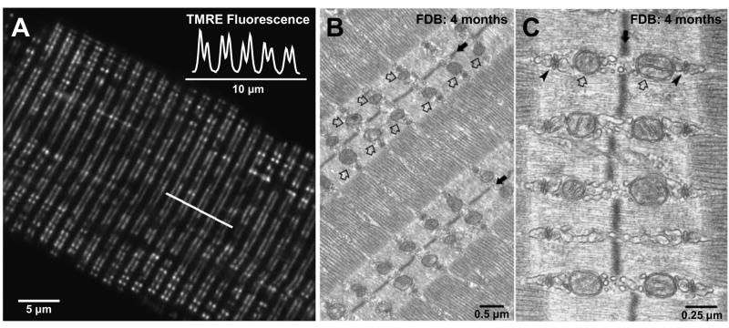
A. Confocal image of a single flexor digitorum brevis (FDB) skeletal muscle fiber obtained from an adult mouse (4 months of age) stained with the mitochondrial-selective dye tetramethylrhodamine ethyl ester (TMRE). TMRE fluorescence along the line of interest marked in A shows characteristic doublets of fluorescence with a sarcomeric periodicity of ~2 μm (inset). B. and C. Representative low (B) and high (C) resolution electron micrographs of FDB skeletal muscle fibers obtained from an adult mouse (4 months of age). Mitochondria (open arrows) are aligned adjacent to the triad (arrowheads), on either size of the Z-line (solid arrows). Images kindly provided by Drs. Simona Boncompagni and Feliciano Protasi.
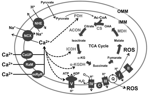
Elevations of calcium (Ca2+) in the mitochondrial matrix stimulate pyruvate dehydrogenase (PDH), isocitrate dehydrogenase (ICDH), α-ketoglutarate dehydrogenase (α-KGDH), and the adenosine triphosphate (ATP) synthetic activity of the F1F0-ATPase (complex V). Low levels of superoxide anions (reactive oxygen species, ROS) are generated as byproducts of electrons being passed to molecular oxygen (O2) at complex I and during the Q cycle (Q). OAA, oxaloacetate; Ac-CoA, acetyl coenzyme A; CS, citrate synthase; ACON, aconitase; α-KG, α-ketoglutarate; MDH, malate dehydrogenase; H+, hydrogen; I to V, complexes I to V; C, cytochrome c; ADP, adenosine diphosphate; H2O, water; OMM, outer mitochondrial membrane; IMM, inner mitochondrial membrane; Na+, sodium; NHE, sodium-hydrogen exchanger; NCX, sodium-calcium exchanger; CaUP, calcium uniporter; RaM; rapid mode calcium transporter; mRyR, mitochondrial ryanodine receptor.
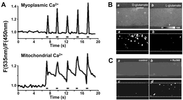
A. Tibialis anterior muscles expressing myoplasmic and mitochondrial calcium (Ca2+)-sensitive cameleons were imaged in vivo with two-photon microscopy. Time course of myoplasmic (top) and mitochondrial (bottom) Ca2+ transients (fluorescence, F) elicited during high frequency stimulation (0.5 s, 50 Hz stimulation train). (Reprinted from Rudolf R, Mongillo M, Magalhaes PJ, Pozzan T. In vivo monitoring of Ca2+ uptake into mitochondria of mouse skeletal muscle during contraction. J Cell Biol. 2004;166(4):527–36. Copyright © 2004 Rockefeller University Press. Used with permission). B. (Upper) Confocal images obtained for permeabilized extensor digitorum longus (EDL) muscle fibers perfused with L-glutamate (right), a substrate for the TCA cycle, or D-glutamate (left), which is not a substrate for the tricarboxylic acid (TCA) cycle. (Lower) Corresponding cumulative binary images for regions with F/F0 > 3 S.D. Data reproduced with permission from J Physiol 2003;547(Pt 2):453–62 (16). C. (Upper) Confocal images obtained for a permeabilized EDL muscle fiber perfused with L-glutamate before (left) and after block of mitochondrial Ca2+ uptake with 20 μM Ru360 (right). (Lower) Corresponding cumulative binary images for regions with F/F0 > 3 S.D. (Reprinted from Isaeva EV, Shirokova N. Metabolic regulation of Ca2+ release in permeabilized mammalian skeletal muscle fibres. J Physiol. 2003;547(Pt 2):453–62. Copyright © 2003 Blackwell Publishing. Used with permission.)
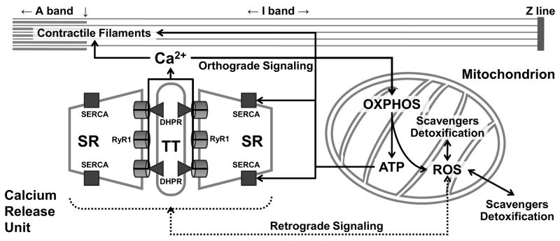
Orthograde sarcoplasmic reticulm (SR)-mitochondrial signaling (solid lines) involves calcium (Ca2+) release during excitation-contraction (EC) coupling being taken up into adjacent mitochondria to stimulate oxidative phosphorylation (OXPHOS) and adenosine triphosphate (ATP) production. Retrograde mitochondrial-SR signaling (broken lines) involves the influence of mitochondrial reactive oxygen species (ROS) production and scavenging/detoxification on the local redox environment and local Ca2+ spark activity of the Ca2+ release unit (CRU). SR, sarcoplasmic reticulum; RyR1, type 1 ryanodine receptor; DHPR, dihydropyridine receptor; TT, transverse tubule; SERCA, sarco(endo)plasmic reticulum Ca2+-ATPase.
Similar articles
-
Sarcoplasmic reticulum-mitochondrial through-space coupling in skeletal muscle.Appl Physiol Nutr Metab. 2009 Jun;34(3):389-95. doi: 10.1139/H09-044. Appl Physiol Nutr Metab. 2009. PMID: 19448704 Free PMC article.
-
Activation and propagation of Ca2+ release from inside the sarcoplasmic reticulum network of mammalian skeletal muscle.J Physiol. 2014 Sep 1;592(17):3727-46. doi: 10.1113/jphysiol.2014.274274. Epub 2014 Jun 27. J Physiol. 2014. PMID: 24973406 Free PMC article.
-
Physical and Functional Cross Talk Between Endo-Sarcoplasmic Reticulum and Mitochondria in Skeletal Muscle.Antioxid Redox Signal. 2020 Apr 20;32(12):873-883. doi: 10.1089/ars.2019.7934. Epub 2019 Dec 11. Antioxid Redox Signal. 2020. PMID: 31825235 Review.
-
Preserved Ca2+ handling and excitation-contraction coupling in muscle fibres from diet-induced obese mice.Diabetologia. 2020 Nov;63(11):2471-2481. doi: 10.1007/s00125-020-05256-8. Epub 2020 Aug 25. Diabetologia. 2020. PMID: 32840676
-
Impaired calcium release during fatigue.J Appl Physiol (1985). 2008 Jan;104(1):296-305. doi: 10.1152/japplphysiol.00908.2007. Epub 2007 Oct 25. J Appl Physiol (1985). 2008. PMID: 17962573 Review.
Cited by
-
Growth Hormone Secretagogues and the Regulation of Calcium Signaling in Muscle.Int J Mol Sci. 2019 Sep 5;20(18):4361. doi: 10.3390/ijms20184361. Int J Mol Sci. 2019. PMID: 31491959 Free PMC article. Review.
-
Endurance exercise attenuates juvenile irradiation-induced skeletal muscle functional decline and mitochondrial stress.Skelet Muscle. 2022 Apr 12;12(1):8. doi: 10.1186/s13395-022-00291-y. Skelet Muscle. 2022. PMID: 35414122 Free PMC article.
-
Doxorubicin causes lesions in the electron transport system of skeletal muscle mitochondria that are associated with a loss of contractile function.J Biol Chem. 2019 Dec 20;294(51):19709-19722. doi: 10.1074/jbc.RA119.008426. Epub 2019 Nov 5. J Biol Chem. 2019. PMID: 31690631 Free PMC article.
-
Antioxidant Treatment Reduces Formation of Structural Cores and Improves Muscle Function in RYR1Y522S/WT Mice.Oxid Med Cell Longev. 2017;2017:6792694. doi: 10.1155/2017/6792694. Epub 2017 Sep 10. Oxid Med Cell Longev. 2017. PMID: 29062463 Free PMC article.
-
Excitation-contraction coupling in mammalian skeletal muscle: Blending old and last-decade research.Front Physiol. 2022 Sep 2;13:989796. doi: 10.3389/fphys.2022.989796. eCollection 2022. Front Physiol. 2022. PMID: 36117698 Free PMC article. Review.
References
-
- Allen DG, Lamb GD, Westerblad H. Skeletal muscle fatigue: Cellular mechanisms. Physiol Rev. 2008;88(1):287–332. - PubMed
-
- Balaban RS. Cardiac energy metabolism homeostasis: Role of cytosolic calcium. J Mol Cell Cardiol. 2002;34(10):1259–71. - PubMed
-
- Boncompagni S, Protasi F. Tethers: Structural connections between SR and the outer mitochondrial membrane. Biophys J. 2007;92:313a. - PubMed
-
- Brookes PS, Yoon Y, Robotham JL, Anders MW, Sheu SS. Calcium, ATP, and ROS: A mitochondrial love-hate triangle. Am J Physiol Cell Physiol. 2004;287(4):C817–C33. - PubMed
-
- Byrd SK. Alterations in the sarcoplasmic reticulum: A possible link to exercise-induced muscle damage. Med Sci Sports Exerc. 1992;24(5):531–6. - PubMed
Publication types
MeSH terms
Substances
Grants and funding
LinkOut - more resources
Full Text Sources
Research Materials
