. 2009 Feb;10(2):186-91.
doi: 10.1038/embor.2008.228. Epub 2008 Dec 19.

Rng3, a member of the UCS family of myosin co-chaperones, associates with myosin heavy chains cotranslationally

Affiliations

Rng3, a member of the UCS family of myosin co-chaperones, associates with myosin heavy chains cotranslationally

Maria J Amorim et al. EMBO Rep. 2009 Feb.

Abstract

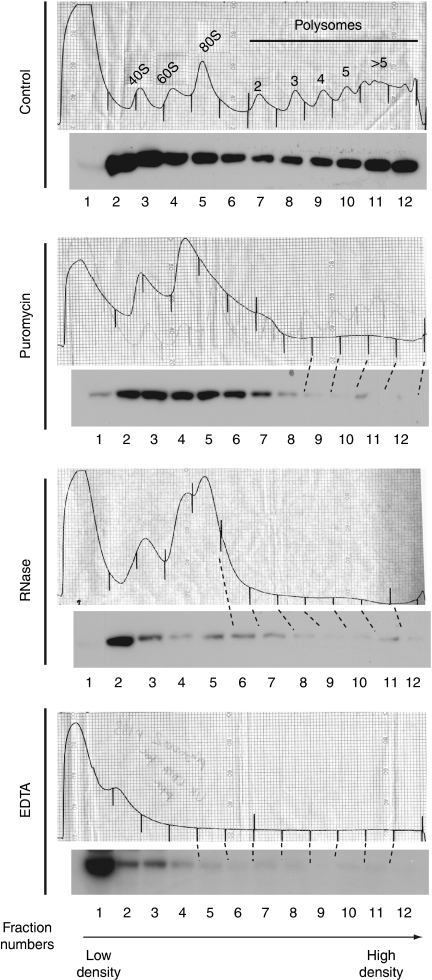
The production of functional myosin heavy chains in many eukaryotic organisms requires the function of proteins containing UCS domains (UNC-45/CRO1/She4), which bind to the myosin head domain and stimulate its folding. UCS proteins are essential for myosin-related functions such as muscle formation, RNA localization and cytokinesis. Here, we show that the Schizosaccharomyces pombe UCS protein Rng3 associates with polysomes, suggesting that UCS proteins might assist myosin folding cotranslationally. To identify Rng3 cotranslational targets systematically, we purified Rng3-associated RNAs and used DNA microarrays to identify the transcripts. Rng3 copurified with only seven transcripts (around 0.1% of S. pombe genes), including all five messenger RNAs encoding myosin heavy chains. These results suggest that every myosin heavy chain in S. pombe is a cotranslational target of Rng3. Furthermore, our data suggest that microarray-based approaches allow the genome-wide identification of cotranslational chaperone targets, and thus pave the way for the dissection of translation-linked chaperone networks.


Conflict of interest statement

The authors declare that they have no conflict of interest.

Figures

Figure 1
Figure 1
Rng3 associates with polysomes. Extracts were fractionated using ultracentrifugation through sucrose gradients and Rng3-TAP was detected by Western blot. The top of the gradient is shown on the left. The position of the ribosomes was determined from the absorbance at 254 nm and confirmed by the distribution of the large subunit ribosomal proteins Rpl7 (data not shown). Ribosomes were stabilized by treatment of cells with cycloheximide, or disassembled by the addition of EDTA, RNase or puromycin to the extracts.
Figure 2
Figure 2
The Rng3 chaperone binds to messenger RNAs encoding myosin heavy chains. (A) Histogram of RNA enrichment ratios from a Rng3-TAP RIp-chip experiment. The x axis represents log2-transformed enrichment ratios that have been median-centred and divided by their standard deviation (that is, the numbers on the axis represent standard deviations above or below the median), and the y axis shows the number of genes in each category. The arrow indicates the threshold used to define enriched RNAs. Only RNAs that were enriched above the threshold in four out of four independent experiments were selected (Table 1). (B) Sections of DNA microarrays hybridized with immunoprecipitated RNAs (magenta) and total RNA (green). The intensity of the signal is proportional to the amount of RNA. The arrows show the position of the myosin RNAs labelled above. TAP only (top row); Rng3-TAP (bottom row). RIp-chip, ribonucleoprotein immunoprecipitation analysed with DNA chips; TAP, tandem affinity purification.
Figure 3
Figure 3
The Rng3–myosin messenger RNA association is sensitive to conditions that destabilize polysomes. (A) Western blots of samples from Rng3-TAP immunoprecipitation experiments from mock-treated (−) or EDTA-treated extracts (+). Here, 1.5% of the input and 5% of the IP were loaded. (B) Enrichment of myosin RNAs in Rng3-TAP IPs (normalized to the levels of the small nuclear RNA U3) from mock- (grey) or EDTA-treated extracts (white). Data from two independent experiments are shown. EX, whole-cell extract; IP, immunoprecipitate; SN, supernatant after immunoprecipitation; TAP, tandem affinity purification.

Similar articles

Cited by

References

    1. Barral JM, Bauer CC, Ortiz I, Epstein HF (1998) Unc-45 mutations in Caenorhabditis elegans implicate a CRO1/She4p-like domain in myosin assembly. J Cell Biol 143: 1215–1225 - PMC - PubMed
    1. Barral JM, Hutagalung AH, Brinker A, Hartl FU, Epstein HF (2002) Role of the myosin assembly protein UNC-45 as a molecular chaperone for myosin. Science 295: 669–671 - PubMed
    1. Bejsovec A, Anderson P (1988) Myosin heavy-chain mutations that disrupt Caenorhabditis elegans thick filament assembly. Genes Dev 2: 1307–1317 - PubMed
    1. Blobel G, Sabatini D (1971) Dissociation of mammalian polyribosomes into subunits by puromycin. Proc Natl Acad Sci USA 68: 390–394 - PMC - PubMed
    1. Chadli A, Graham JD, Abel MG, Jackson TA, Gordon DF, Wood WM, Felts SJ, Horwitz KB, Toft D (2006) GCUNC-45 is a novel regulator for the progesterone receptor/hsp90 chaperoning pathway. Mol Cell Biol 26: 1722–1730 - PMC - PubMed

Publication types

MeSH terms