. 2009 Jan 15;457(7227):277-80.
doi: 10.1038/nature07677. Epub 2008 Dec 21.

Induced pluripotent stem cells from a spinal muscular atrophy patient

Affiliations

Induced pluripotent stem cells from a spinal muscular atrophy patient

Allison D Ebert et al. Nature. .

Abstract

Spinal muscular atrophy is one of the most common inherited forms of neurological disease leading to infant mortality. Patients have selective loss of lower motor neurons resulting in muscle weakness, paralysis and often death. Although patient fibroblasts have been used extensively to study spinal muscular atrophy, motor neurons have a unique anatomy and physiology which may underlie their vulnerability to the disease process. Here we report the generation of induced pluripotent stem cells from skin fibroblast samples taken from a child with spinal muscular atrophy. These cells expanded robustly in culture, maintained the disease genotype and generated motor neurons that showed selective deficits compared to those derived from the child's unaffected mother. This is the first study to show that human induced pluripotent stem cells can be used to model the specific pathology seen in a genetically inherited disease. As such, it represents a promising resource to study disease mechanisms, screen new drug compounds and develop new therapies.


Figures
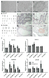
Figure 1
Figure 1
Newly generated iPS cells were fully reprogrammed. a-c, iPS-WT and iPS-SMA cells formed tightly packed colonies in contrast to the spindle morphology of fibroblasts. d,e, No karyotypic abnormalities were observed. Following transplantation, all iPS cells generated teratomas showing f, neural tissue (ectoderm), g, primitive gut (endoderm), h, cartilage (mesoderm), and i, bone (mesoderm). j, Quantitative RT-PCR showed induction of endogenous transcripts of OCT 4, SOX 2, NANOG, and LIN 28. “Endogenous” refers to primers recognizing the 3′ untranslated region, whereas “Total” identifies both the endogenous and exogenously expressed transgene. Data are expressed as mean ± s.e.m. Scale bar = 50μm
Figure 2
Figure 2
iPS-SMA show decreased SMN transcripts and lack of gems. a, iPS-WT and iPS-SMA cells had levels of full-length (FL) and truncated, exon 7 deleted (Δ7) SMN transcripts similar to their respective fibroblast lines (Fib-WT and Fib-SMA, respectively). b, Additionally, both Fib-SMA and iPS-SMA cells showed a specific lack of SMN1, although SMN2 FL and □7 transcripts were still present in all samples. c, Quantitative RT-PCR showed reduced FL-SMA transcript in both fibroblasts (61.1 % ± 0.6 reduced) and iPS cells (67.4 % ± 0.8 reduced) when compared to WT cultures (Student's t-test *p<0.001). Data are presented as mean ± s.e.m.
Figure 3
Figure 3
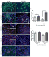
iPS-WT and iPS-SMA cells can generate cells in the neural lineage. a,b, iPS-WT and iPS-SMA cells generated nestin positive neural progenitor cells (green); c,d, Tuj1 positive neurons (green) and GFAP positive astrocytes (red); e,f, HB9 (green)/ChAT (red) double positive motor neurons, and g,h, SMI-32 (red) positive motor neurons at 4-6 weeks of differentiation i, At 8 weeks, punctate synapsin staining (green) on SMI-32 positive motor neurons (red) was identified on iPS-WT cells. j, However, only diffuse synapsin staining (green) was observed on SMI-32 positive motor neurons (red) in iPS-SMA cells. Arrowheads in i and j show punctate synapsin staining on SMI-32 negative cells. Nuclei are labeled with Hoechst nuclear dye (blue). k,l, iPS-SMA-derived motor neurons are significantly reduced in number and size at 6 weeks compared to iPS-WT cells (n=3 ANOVA, * p<0.05). All data are presented as mean ± s.e.m. Scale bar = 50μm (a-h), 25μm (i,j).
Figure 4
Figure 4
iPS-WT and iPS-SMA cells increase SMN protein in response to drug treatment. a,c, Untreated Fib-WT and iPS-WT cells show nuclear gem localization, whereas b,d, untreated Fib-SMA and iPS-SMA lack nuclear gems. f,h,i, Following valproic acid or tobramycin treatment, iPS-SMA cells show a significant increase the number of gems (ANOVA, p<0.05). Gems are indicated by arrows. j,k, Western blot analysis following valproic acid or tobramycin treatment showed a significant 2-3 fold increase in SMN protein in iPS-SMA cells compared to iPS-SMA cells (n=3 ANOVA, *p<0.05; **p<0.01). Data are presented as mean ± s.e.m. Scale bar = 50μm.

Comment in

Similar articles

Cited by

References

    1. Lefebvre S, et al. Identification and characterization of a spinal muscular atrophy-determining gene. Cell. 1995;80:155–165. - PubMed
    1. Coovert DD, et al. The survival motor neuron protein in spinal muscular atrophy. Hum Mol Genet. 1997;6:1205–1214. - PubMed
    1. Crawford TO, Pardo CA. The neurobiology of childhood spinal muscular atrophy. Neurobiol Dis. 1996;3:97–110. - PubMed
    1. Munsat TL, Davies KE. Neuromuscul Disord; International SMA consortium meeting; 26-28 June 1992; Bonn, Germany. 1992. pp. 423–428. - PubMed
    1. Lorson CL, Hahnen E, Androphy EJ, Wirth B. A single nucleotide in the SMN gene regulates splicing and is responsible for spinal muscular atrophy. Proc Natl Acad Sci U S A. 1999;96:6307–6311. - PMC - PubMed

Publication types

MeSH terms

Substances

Associated data