. 2009 Feb;11(2):183-9.
doi: 10.1038/ncb1825. Epub 2008 Dec 21.

UNC-6 (netrin) orients the invasive membrane of the anchor cell in C. elegans

Affiliations

UNC-6 (netrin) orients the invasive membrane of the anchor cell in C. elegans

Joshua W Ziel et al. Nat Cell Biol. 2009 Feb.

Abstract

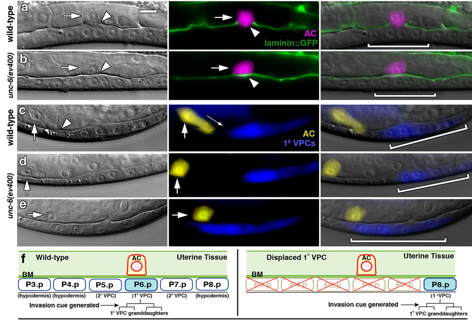
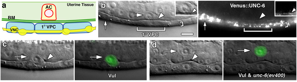
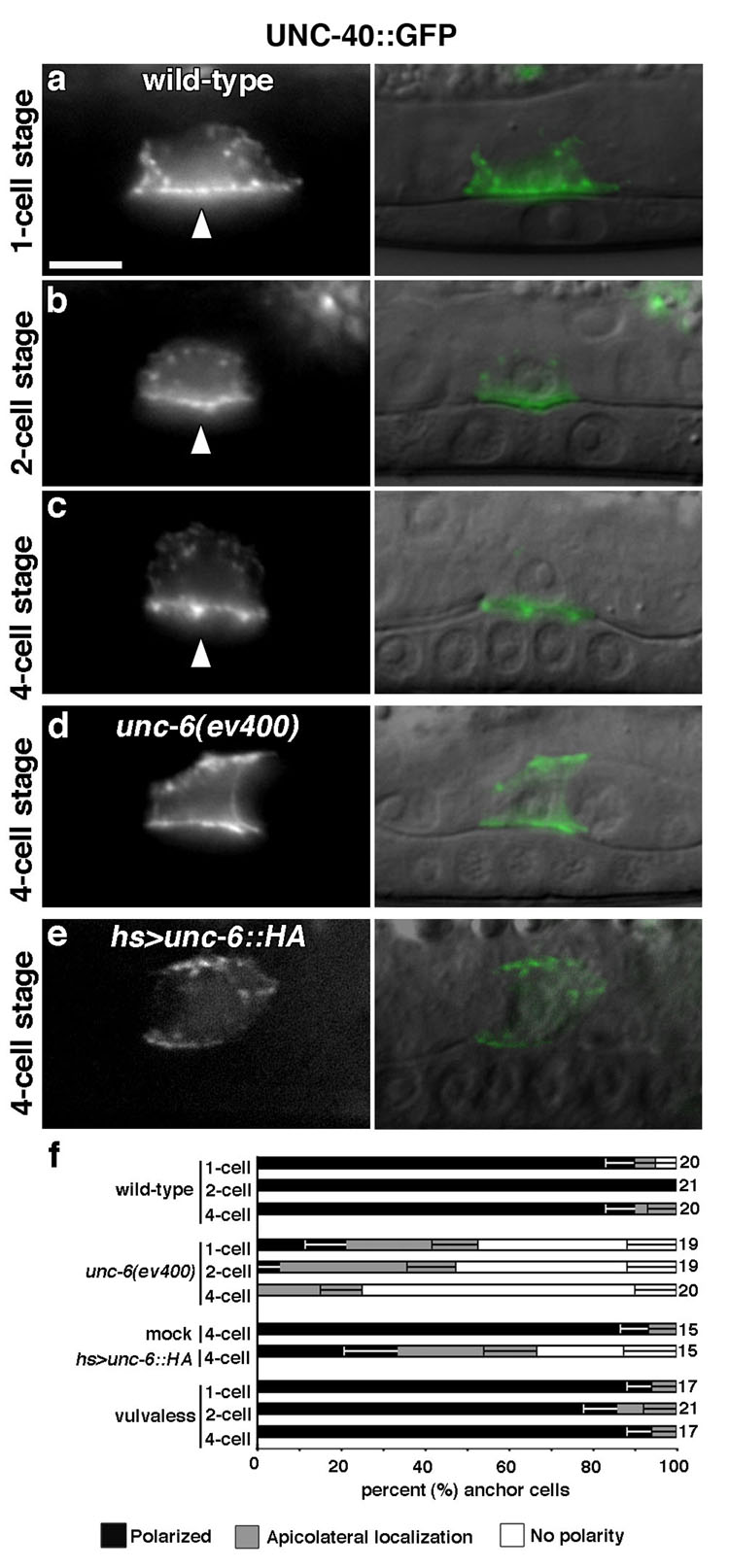
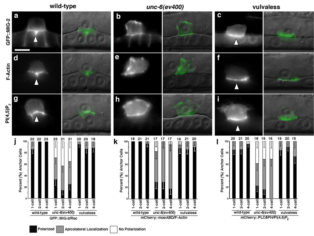
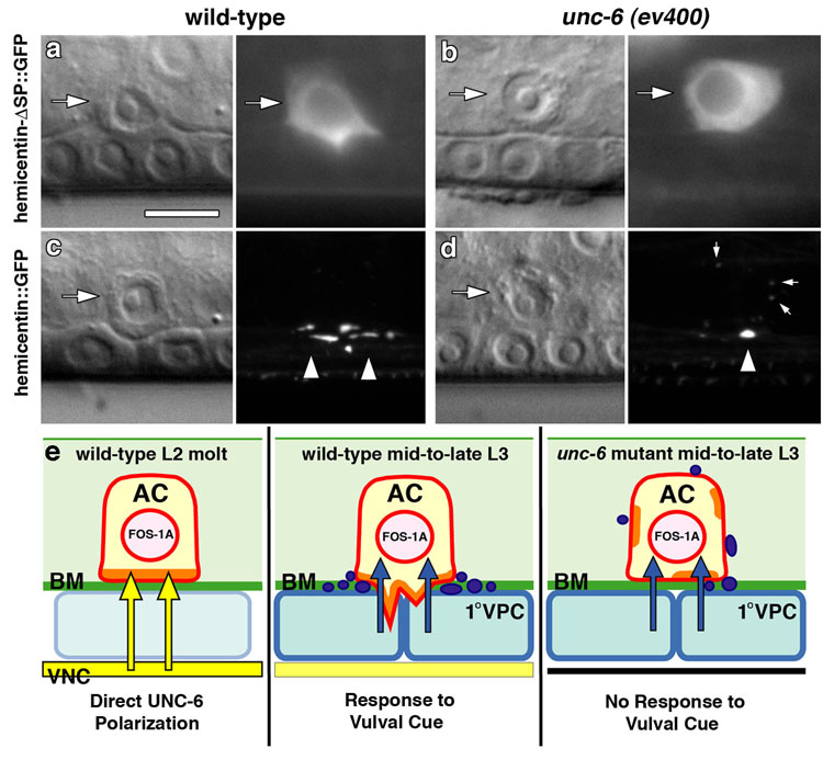
Despite their profound importance in the development of cancer, the extracellular cues that target cell invasion through basement membrane barriers remain poorly understood. A central obstacle has been the difficulty of studying the interactions between invading cells and basement membranes in vivo. Using the genetically and visually tractable model of Caenorhabditis elegans anchor cell (AC) invasion, we show that UNC-6 (netrin) signalling, a pathway not previously implicated in controlling cell invasion in vivo, is a key regulator of this process. Site of action studies reveal that before invasion, localized UNC-6 secretion directs its receptor, UNC-40, to the plasma membrane of the AC, in contact with the basement membrane. There, UNC-40 polarizes a specialized invasive membrane domain through the enrichment of actin regulators, F-actin and phosphatidylinositol 4,5-bisphosphate (PtdIns(4,5)P(2)). Cell ablation experiments indicate that UNC-6 promotes the formation of invasive protrusions from the AC that break down the basement membrane in response to a subsequent vulval cue. Together, these results characterize an invasive membrane domain in vivo, and reveal a role for UNC-6 (netrin) in polarizing this domain towards its basement membrane target.


Figures

Figure 1
Figure 1. The AC fails to invade in unc-6 (netrin) mutants
Late L3 animals; anterior left, ventral down; bracket, 1° VPCs. (a) Nomarski (left), fluorescence (center), and overlaid images (right) show that the wild-type AC (arrows; expressing zmp-1>mCherry in magenta) has crossed basement membrane (arrowhead; interruption of phase dense line on left and basement membrane component laminin, LAM-1∷GFP in green) and contacted the central 1° fated P6.p granddaughters (P6.p 4-cell stage; 20/20 animals). (b) In unc-6 mutants, the basement membrane was intact under the AC in most animals (18/21 animals), and showed only small gaps in those that had partially invaded (3/21 animals, not shown). (c) In wild-type animals the AC (expressing cdh-3>YFP in yellow) extended invasive processes toward isolated 1° fated P8.p cell descendants (expressing egl-17>CFP in blue) in all cases examined (up to 25µm away; 42/42 animals). (d,e) In unc-6 mutants the AC failed to extend invasive processes toward 1° fated P8.p cell descendants (44/44 animals), even when they directly bordered the AC (e, 9/9 animals). (f) Schematic diagram showing the AC in wild-type animals at the early L3 stage (P6.p 1-cell stage, left), and after ablation of the VPCs P3.p through P7.p (right). Both diagrams show animals prior to division of the 1° fated VPC (shown in blue). Future divisions of the 1° VPC prior to and during invasion are shown below in brackets. The arrow points to the time that the 1° VPC cue is generated that stimulates invasion. At this time the AC breaks through the underlying gonadal and ventral epidermal basement membranes (BM) and invades towards the 1° VPCs. The scale bar (upper left panel) is 5 µm for this and all other figures.
Figure 2
Figure 2. UNC-6 is a distinct VNC-derived pro-invasive cue
(a) A schematic image along the left side of the animal showing the relationship of the AC, basement membrane (BM), 1° VPC, and the neighboring ventral nerve cord (VNC) with associated cell bodies (yellow ovals). (b) Nomarski (left) and fluorescence (right) images of an animal expressing Venus∷UNC-6 viewed at the focal plane between the 1° VPC (large bracket) and the VNC. Venus∷UNC-6 accumulated in the VNC (small bracket, small arrow points to VNC cell body where accumulation was strongest), and low levels were found in the basement membrane underlying the AC (arrowheads). Insets show an enlarged image of the AC with Venus∷UNC-6 localization ventrally in the basement membrane (arrowheads). (c) In approximately 20% of vulvaless animals the AC invades into the underlying epidermis (both in lin-3 loss of function mutants or when removed by laser ablation; Table S2). A Nomarski image (left) and a cdh-3>GFP overlay (right) show an AC in a vulvaless animal (arrow) that has broken through the underlying basement membrane and invaded (arrowhead). (d) In vulvaless animals carrying the unc-6 mutation, the AC never invaded (70/70 animals) and usually detached from the basement membrane at the early L4 stage (arrowhead; 65/70 animals; Table S2).
Figure 3
Figure 3. UNC-6 directs its receptor UNC-40 to the invasive cell membrane
Fluorescence image (left), Nomarski overlay (right). (a-c) UNC-40∷GFP was present within intracellular vesicles and polarized to the AC’s invasive cell membrane (arrowheads) during the late L2 molt (P6.p 1-cell stage; 5–6 hours prior to invasion). UNC-40∷GFP maintained this polarization until the time of invasion at the P6.p 4-cell stage. (d) UNC-40∷GFP polarization was perturbed in unc-6 mutants, and (e) in wild-type animals ubiquitously expressing UNC-6∷HA induced by heat shock. (f) Compared with wild-type controls, UNC-40∷GFP polarization in unc-6 mutants, or following ubiquitous UNC-6∷HA expression was significantly perturbed (P < 7×10⁻⁴ in all cases, Fisher’s exact test in this and subsequent graphs). In contrast, neither vulvaless nor mock heat shocked animals showed changes in UNC-40∷GFP polarity compared with wild-type (P > 0.05). The number of animals examined at each stage is listed to the right of the graph; error bars report the standard error of the proportion.
Figure 4
Figure 4. UNC-6 localizes the Rac protein MIG-2, F-actin, and PI(4,5)P2 to the invasive cell membrane
Fluorescence image (left), Nomarski overlaid image (right). (a, d, g) The Rac protein GFP∷MIG-2, the F-actin binding protein mCherry∷moeABD and the phosphatidylinositol 4,5-bisphosphate sensor mCherry∷PLCδPH were localized to the invasive cell membrane in wild-type animals (arrowheads). The endogenous mig-2 promoter also drove low levels of expression in the vulval cells. (b, e, h) In unc-6 mutants, MIG-2, F-actin and PI(4,5)P2 failed to polarize to the invasive cell membrane. (c, f, i) In contrast, MIG-2, F-actin and PI(4,5)P2 were polarized normally in vulvaless animals (arrowheads). (j, k, l) Quantification of MIG-2, F-actin and PI(4,5)P2 polarization, respectively, in wild-type, unc-6, and vulvaless animals prior to and during invasion. Localization of all markers was significantly perturbed in unc-6 animals at all time points examined compared to wild-type controls (P >0.0003 in all cases). In contrast, vulvaless animals showed no significant changes in polarity (P > 0.05). The number of animals examined at each stage is noted at the top of each bar; error bars show the standard error of the proportion and raw percentages are reported in Table S3.
Figure 5
Figure 5. UNC-6 promotes the deposition of hemicentin at the site of invasion
Nomarski images (left) and corresponding fluorescence images (right). (a) Expression of the transcriptional reporter hemicentin-ΔSP∷GFP within the AC (arrow) in wild-type animals during invasion. (b) Hemicentin-ΔSP∷GFP was expressed at the same levels in unc-6 mutants (n = 20 for each; P = 0.69, unpaired t-test). (c) Full length hemicentin∷GFP is deposited under the AC’s invasive membrane (arrowheads) during invasion. (d) In unc-6 mutants, there was a 65% reduction in hemicentin deposition under the invasive cell membrane (arrowhead; P = 5.0×10⁻⁶, unpaired t-test), and a three fold increase in accumulations formed along apical and lateral membranes (small arrows; P = 0.022, unpaired t-test, n = 18 for each; Videos S1, S2). We found no significant correlation (correlation coefficient = −0.248, P = 0.320 Students t-test, df = 16) between perturbations in hemicentin deposition and the contact area with the basement membrane. (e) Schematic diagram of the role of unc-6 (netrin) in regulating AC invasion. During the L2 molt, UNC-6 protein secreted from the ventral nerve cord (VNC and arrows in yellow) polarizes its receptor UNC-40 to the AC’s plasma membrane in contact with the basement membrane (BM, green). There, UNC-40 establishes a specialized invasive membrane domain (orange) containing F-actin, and its effectors--actin regulators and the phospholipid PI(4,5)P2. Approximately 4–6 hours later, hemicentin is deposited under the invasive cell membrane (puncta, purple) and invasive protrusions are generated in response to the 1° vulval cell cue (VPCs, arrows in blue). In unc-6 mutants, the invasive membrane is not polarized, hemicentin deposition is reduced and mistargeted, and the AC fails to generate invasive processes in response to the 1° VPC signal. Importantly, this model for UNC-40 localization does not preclude the possibility of feed-back mechanisms that regulate UNC-40 localization and function.

References

    1. Machesky L, Jurdic P, Hinz B. Grab, stick, pull and digest: the functional diversity of actin-associated matrix-adhesion structures. Workshop on Invadopodia, Podosomes and Focal Adhesions in Tissue Invasion. EMBO Rep. 2008;9:139–143. - PMC - PubMed
    1. Even-Ram S, Yamada KM. Cell migration in 3D matrix. Curr Opin Cell Biol. 2005;17:524–532. - PubMed
    1. Hotary K, Li XY, Allen E, Stevens SL, Weiss SJ. A cancer cell metalloprotease triad regulates the basement membrane transmigration program. Genes Dev. 2006;20:2673–2686. - PMC - PubMed
    1. Sharma-Kishore R, White JG, Southgate E, Podbilewicz B. Formation of the vulva in Caenorhabditis elegans: a paradigm for organogenesis. Development. 1999;126:691–699. - PubMed
    1. Sherwood DR, Sternberg PW. Anchor cell invasion into the vulval epithelium in C. elegans. Dev Cell. 2003;5:21–31. - PubMed

Publication types

MeSH terms