

High-resolution structure of the open NaK channel
- PMID: 19098917
- PMCID: PMC2615073
- DOI: 10.1038/nsmb.1531
High-resolution structure of the open NaK channel
Abstract
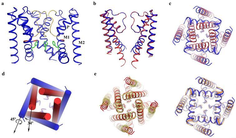
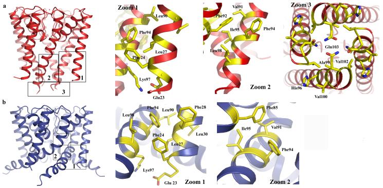
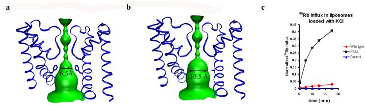
We report the crystal structure of the nonselective cation channel NaK from Bacillus cereus at a resolution of 1.6 A. The structure reveals the intracellular gate in an open state, as opposed to the closed form reported previously, making NaK the only channel for which the three-dimensional structures of both conformations are known. Channel opening follows a conserved mechanism of inner helix bending using a flexible glycine residue, the gating hinge, seen in MthK and most other tetrameric cation channels. Additionally, distinct inter and intrasubunit rearrangements involved in channel gating are seen and characterized for the first time along with inner helix twisting motions. Furthermore, we identify a residue deeper within the cavity of the channel pore, Phe92, which is likely to form a constriction point within the open pore, restricting ion flux through the channel. Mutating this residue to alanine causes a subsequent increase in ion-conduction rates as measured by (86)Rb flux assays. The structures of both the open and closed conformations of the NaK channel correlate well with those of equivalent K(+) channel conformations, namely MthK and KcsA, respectively.
Figures
References
-
- Hille B. Ion Channels of Excitable Membranes. 3rd edition ed. Sinauer Associates, Inc.; Sunderland, MA: 2001.
-
- Jiang Y, et al. Crystal structure and mechanism of a calcium-gated potassium channel. Nature. 2002;417(6888):515–22. - PubMed
-
- Zhou Y, et al. Chemistry of ion coordination and hydration revealed by a K+ channel-Fab complex at 2.0 A resolution. Nature. 2001;414(6859):43–8. - PubMed
-
- Jiang Y, et al. The open pore conformation of potassium channels. Nature. 2002;417(6888):523–6. - PubMed
-
- Doyle DA, et al. The structure of the potassium channel: molecular basis of K+ conduction and selectivity. Science. 1998;280(5360):69–77. - PubMed
Publication types
MeSH terms
Substances
Associated data
- Actions
Grants and funding
LinkOut - more resources
Full Text Sources
Molecular Biology Databases
Miscellaneous
