. 2009 Feb;10(2):167-75.
doi: 10.1038/ni.1690. Epub 2008 Dec 21.

The costimulatory molecule ICOS regulates the expression of c-Maf and IL-21 in the development of follicular T helper cells and TH-17 cells

Affiliations

The costimulatory molecule ICOS regulates the expression of c-Maf and IL-21 in the development of follicular T helper cells and TH-17 cells

Aurelie T Bauquet et al. Nat Immunol. 2009 Feb.

Abstract

The inducible costimulatory molecule ICOS has been suggested to be important in the development of interleukin 17 (IL-17)-producing helper T cells (T(H)-17 cells) and of follicular helper T cells (T(FH) cells). Here we show that ICOS-deficient mice had no defect in T(H)-17 differentiation but had fewer T(H)-17 cells after IL-23 stimulation and fewer T(FH) cells. We also show that T(FH) cells produced IL-17 and that T(FH) cells in ICOS-deficient mice were defective in IL-17 production. Both T(H)-17 and T(FH) cells had higher expression of the transcription factor c-Maf. Genetic loss of c-Maf resulted in a defect in IL-21 production and fewer T(H)-17 and T(FH) cells. Thus our data suggest that ICOS-induced c-Maf regulates IL-21 production that in turn regulates the expansion of T(H)-17 and T(FH) cells.


Figures

Figure 1
Figure 1. ICOS is not crucial for T -17 differentiation in vitro
(a,b) Naïve CD4+CD62Lhigh T cells from ICOS-deficient (Icos-/-) or wild-type mice were stimulated with soluble anti-CD3, antigen-presenting cells, IL-6 and TGF-β for 5 days and then were restimulated with PMA-ionomycin. IL-17 production in CD4+ cells was measured by intracellular cytokine staining (a) or by ELISA (b). Numbers adjacent to boxed areas or in quadrants indicate percent positive cells in each quadrant. Data represent the mean ± s.d. of six experiments (a) or one of four experiments (b) (mean ± s.d. of triplicate wells).
Figure 2
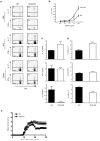
Figure 2. ICOS is not crucial for TH-17 differentiation in vivo nor for induction of acute EAE
(a-e) ICOS-deficient (Icos-/-) or wild-type mice were immunized with MOG35-55 emulsified in complete Freund's adjuvant to induce EAE. (a) Flow cytometry for intracellular cytokine secretion in CD4+ T cells at day 10 obtained from mononuclear cells isolated from CNS, dLN and spleen activated with PMA-ionomycin. (b-d) Lymph node cells from immunized mice were tested for antigen-specific response: proliferation in triplicate cultures by [H] thymidine incorporation (mean ± s.d.) (b), cytokine production by ELISA (day 2) (c) and intracellular cytokine staining by flow cytometry (day 5) (d). (e) The mean clinical EAE scores for the ICOS-deficient (dark circles) and wild-type control (open circles) groups (mean ± s.e.m.). Numbers adjacent to boxed areas or in quadrants indicate percent positive cells in each. Data represent one of four experiments (a) or the mean ± s.d. of five (b,c,d) or four (e) experiments.
Figure 3
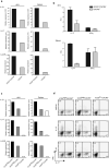
Figure 3. TFH cells produce IL-17
(a-d) FACS-sorted CD4+ICOShiCXCR5+, CD4+ICOShiCXCR5- and CD4+ICOSloCXCR5- T cells were sorted by flow cytometry from wild-type immunized mice and then analyzed. (a) RT-PCR for expression of IL-21, IL-17 and IL-23R in CD4+ICOShiCXCR5+ (black histograms) and (CD4+CXCR5-) light grey histograms) T cells from draining lymph node or spleen 4 hr after restimulation of the cells in vitro with PMA/ionomycin; expression relative to β-actin. (b) Flow cytometry for expression of IL-17 and IFN-γ by cells in a stimulated instead with soluble anti-CD3, antigen-presenting cells and IL-23 for 4 days. (c) RT-PCR for expression of IL-21, IL-17 and IL-23R mRNA (relative to β-actin) in the indicated cells after treatment with PMA-ionomycin for 4 hours. (d) Flow cytometry of the indicated cells stimulated in vitro with IL-12 or IL-23 for 4 days and then restimulated with PMA-ionomycin. Numbers adjacent to boxed areas or in quadrants indicate percent positive cells in each. Data represent one of six experiments (a) or mean ± s.d. of three experiments (b).
Figure 4
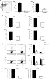
Figure 4. ICOS-deficient TFH cells are defective in IL-17 production
(a-c) CD4+CXCR5+ from dLN and spleens of wild-type and ICOS-deficient immunized mice were restimulated with PMA-ionomycin for 4 hours. The percentage of CD4+CXCR5+ cells was assessed by flow cytometry (a) and the expression of IL-21 (b) and IL-17 (c) was assessed by quantitative PCR relative. (d) Flow cytometry of CD4+CXCR5+ cells from wild-type or ICOS-deficient immunized mice were stimulated with anti-CD3, antigen-presenting cells and IL-23 for 4 days and then restimulated with PMA-ionomycin and the percentage of IL-17+ and IFN-γ+ cells was measured by intracellular cytokine staining. (e) RT-PCR for IL-23R expression in CD4+CXCR5+ cells from dLN and spleens of wild-type and ICOS-deficient immunized mice restimulated with PMA-ionomycin for 4 hours; expression is relative to β-actin. Numbers adjacent to boxed areas or in quadrants indicate percent positive cells in each. Data represent the mean ± s.d. of four (a) or three (d) experiments or represent one of six (b,c) or four (e) experiments.
Figure 5
Figure 5. ICOS-deficient T cells show a defect in secondary TH-17 responses
(a-c) Flow cytometry of ICOS-deficient or wild-type CD4+ T cells (left panel) or CD4+CD62Llo T cells (right panel) stimulated with anti-CD3, APCs, and IL-23 for 5 days and then restimulated with PMA-ionomycin (a); wild-type or ICOS-deficient cells differentiated for 5 days with IL-6 and TGF-β for 5 days and then restimulated with anti-CD3, APCs and IL-23 for an additional 4 days (b); or lymph nodes cells from immunized wild-type or ICOS-deficient mice cultured with MOG35-55 and IL-23 for 4 days (c). (d,e) RT-PCR expression of IL-12Rβ1 and IL-23R (d) and ELISA for production of IL-21 (e) from naïve T cells from ICOS-deficient or wild-type mice stimulated with soluble anti-CD3, APCs, IL-6 and TGF-β for 5 days; expression to β-actin. Data represent the mean ± s.d. of four (a,b) experiments or represent one of three (c) or two (d,e) experiments.
Figure 6
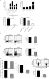
Figure 6. c-Maf in TH-17 differentiation
(a) RT-PCR of c-Maf expression in CD4+CD62Lhigh T cells from wild-type mice stimulated under TH0, TH1, TH2, TH-17 conditions, or as indicated with plate bound anti-CD3 and anti-CD28 (top left) or with anti-CD3 and APCs (top right) (expression relative to GADPH); c-Maf expression in ICOS-deficient or wild-type naïve CD4+CD62Lhi T cells stimulated with anti-CD3, antigen-presenting cells, IL-6 and TGF-β (bottom left; expression relative to β-actin); c-Maf expression in CD4+ICOShiCXCR5+, CD4+ICOShiCXCR5- and CD4+ICOSloCXCR5- cells from dLN of wild-type mice immunized with MOG and restimulated in vitro; (bottom right; expression relative to β-actin). (b,c) Flow cytometry (left) and ELISA (right) for IFN-γ and IL-17 production by c-Maf-deficient or wild-type naïve CD4+CD62Lhi T cells stimulated in vitro (b) or c-Maf-deficient or wild-type TH-17 cells restimulated in vitro (c). (d) RT-PCR for IL-23R expression in TH-17 cells day 3 of ‘primary differentiation’ (TGF-β + IL-6; left) or ‘secondary expansion’ (+IL-23, right); expression is relative to β-actin. (e) ELISA for IL-21 production at day 3 of primary differentiation of TH-17. (f) Flow cytometry of wild-type and c-Maf-deficient mice immunized with TNP-OVA in complete Freund's adjuvant. Numbers indicate percent positive TFH cells (CD4+CXCR5+ICOS+) in each quadrant.

Comment in

References

    1. Lock C, et al. Gene-microarray analysis of multiple sclerosis lesions yields new targets validated in autoimmune encephalomyelitis. Nat Med. 2002;8:500–508. - PubMed
    1. Matusevicius D, et al. Interleukin-17 mRNA expression in blood and CSF mononuclear cells is augmented in multiple sclerosis. Mult Scler. 1999;5:101–104. - PubMed
    1. Aarvak T, Chabaud M, Miossec P, Natvig JB. IL-17 is produced by some proinflammatory Th1/Th0 cells but not by Th2 cells. J Immunol. 1999;162:1246–1251. - PubMed
    1. Teunissen MB, Koomen CW, de Waal Malefyt R, Wierenga EA, Bos JD. Interleukin-17 and interferon-gamma synergize in the enhancement of proinflammatory cytokine production by human keratinocytes. J Invest Dermatol. 1998;111:645–649. - PubMed
    1. Langrish CL, et al. IL-23 drives a pathogenic T cell population that induces autoimmune inflammation. J Exp Med. 2005;201:233–240. - PMC - PubMed

Publication types

MeSH terms