. 2008;3(12):e3989.
doi: 10.1371/journal.pone.0003989. Epub 2008 Dec 22.

Role of Cajal bodies and nucleolus in the maturation of the U1 snRNP in Arabidopsis

Affiliations

Role of Cajal bodies and nucleolus in the maturation of the U1 snRNP in Arabidopsis

Zdravko J Lorković et al. PLoS One. 2008.

Abstract

Background: The biogenesis of spliceosomal snRNPs takes place in both the cytoplasm where Sm core proteins are added and snRNAs are modified at the 5' and 3' termini and in the nucleus where snRNP-specific proteins associate. U1 snRNP consists of U1 snRNA, seven Sm proteins and three snRNP-specific proteins, U1-70K, U1A, and U1C. It has been shown previously that after import to the nucleus U2 and U4/U6 snRNP-specific proteins first appear in Cajal bodies (CB) and then in splicing speckles. In addition, in cells grown under normal conditions U2, U4, U5, and U6 snRNAs/snRNPs are abundant in CBs. Therefore, it has been proposed that the final assembly of these spliceosomal snRNPs takes place in this nuclear compartment. In contrast, U1 snRNA in both animal and plant cells has rarely been found in this nuclear compartment.

Methodology/principal findings: Here, we analysed the subnuclear distribution of Arabidopsis U1 snRNP-specific proteins fused to GFP or mRFP in transiently transformed Arabidopsis protoplasts. Irrespective of the tag used, U1-70K was exclusively found in the nucleus, whereas U1A and U1C were equally distributed between the nucleus and the cytoplasm. In the nucleus all three proteins localised to CBs and nucleoli although to different extent. Interestingly, we also found that the appearance of the three proteins in nuclear speckles differ significantly. U1-70K was mostly found in speckles whereas U1A and U1C in approximately 90% of cells showed diffuse nucleoplasmic in combination with CBs and nucleolar localisation.

Conclusions/significance: Our data indicate that CBs and nucleolus are involved in the maturation of U1 snRNP. Differences in nuclear accumulation and distribution between U1-70K and U1A and U1C proteins may indicate that either U1-70K or U1A and U1C associate with, or is/are involved, in other nuclear processes apart from pre-mRNA splicing.


Conflict of interest statement

Competing Interests: The authors have declared that no competing interests exist.

Figures

Figure 1
Figure 1. Localisation of transiently expressed U1 snRNP proteins in Arabidopsis protoplasts.
(A) Single confocal sections of protoplasts expressing U1-70K, U1A, and U1C proteins fused to GFP. Corresponding differential interference contrast (DIC) image of a cell expressing U1-70K is also shown. Arrows, broken arrow and arrowheads point to nuclei, nucleoli and CBs, respectively. Scale bars, 15 µm. (B) Cellular localisation of GFP- (left panel) or HA-tagged (right panel) U1 snRNP-specific proteins studied by cellular fractionation. Cell extracts were fractionated as described . Lanes T, C, and N; total cellular, cytoplasmic, and nuclear protein fractions, respectively. Proteins were resolved by SDS-PAGE and analyzed by Western blotting, using mouse anti-GFP and rat anti-HA mAb. Molecular mass standards in kDa are indicated on the left. To control the quality of the fractionation procedure the same blots were probed with antibodies against nuclear and cytoplasmic proteins RBP45 and fructose 1,6-bisphosphatase (cFBP), respectively (two bottom panels). (C) Immunodetection of U1-70K, U1A and U1C GFP fusion proteins in protein extract from transformed protoplasts. Total protein extracts were analysed by SDS-PAGE and Western blotting with anti-GFP antibody. Molecular mass standards in kDa are indicated on the left. Western blotting of the same protein extracts with anti-tubulin antibodies was performed as a loading control (bottom panel).
Figure 2
Figure 2. Transiently expressed U1 snRNP-specific proteins assemble into mature snRNP.
(A) Immunoprecipitation of U1A-GFP and U1C-GFP fusion proteins with anti-m3G antibody (α-m3G). Lanes 1, input protein extract. Lanes 2, protein extracts incubated with protein-A Sepharose (pA). Lanes 3, immunoprecipitations with anti-m3G antibody (α-m3G). Arrowheads and arrows point to precipitated proteins and immunoglobulin heavy chains, respectively. The blot was probed with anti-GFP antibody. (B) Immunoprecipitation of U1A-HA and U1C-HA fusion proteins with anti-m3G antibody (α-m3G). The blots were probed with anti-HA antibody. Other details as in (A). (C) U1A-GFP and U1C-GFP fusion proteins precipitated with anti-GFP antibody co-immunoprecipitate U1 snRNAs. Left panel: lane 1, immunoprecipitation with anti-GFP antibody with protein extract from non-transformed protoplasts; lanes 2 and 5, input protein extract from cells expressing U1A-GFP and U1C-GFP fusion proteins, respectively; lanes 3 and 6, protein extracts from transformed cells incubated with protein-A Sepharose only (pA); lanes 4 and 7, immunoprecipitations with anti-GFP antibody (α-GFP) with protein extracts from transformed protoplasts. Arrowheads point to U1A and U1C GFP-tagged proteins and arrows point to immunoglobulin heavy and light chains. Right panel: analysis of anti-GFP immunoprecipitates (from the left panel, lanes 1, 4, and 7) for the presence of U1 snRNAs. After immunoprecipitation RNA was extracted, labelled by [32P]-pCp ligation and analyzed on 8% denaturing PAA gels. Lane 1, RNA immunoprecipitated with anti-GFP antibody from non-transformed cells. Lanes 4 and 7, RNA co-precipitated with U1A-GFP and U1C-GFP, respectively.
Figure 3
Figure 3. Nuclear distribution of transiently expressed U1-70K, U1A, and U1C proteins.
Representative images of nuclear patterns observed in protoplasts expressing U1-70K (A), U1A (B), and U1C (C) GFP-tagged proteins. Single confocal sections are shown. Arrows, arrowheads, and asterisks point to nucleoli, CBs, and nucleolar cavities, respectively. Scale bars, 8 µm.
Figure 4
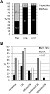
Figure 4. Quantification of nuclear patterns shown in Figure 3.
Transformations with GFP tagged proteins were performed independently three times, and each time 100 randomly chosen cells were analysed. (A) Speckled and diffuse nucleoplasmic staining patterns were scored and percentages are indicated inside the bars. (B) CB and nucleolar localisation as well as combinations of different patterns as indicated on X-axis were scored. The Y-axis represents the percentage of cells showing specific pattern(s).
Figure 5
Figure 5. Co-localisation studies with U1 snRNP-specific proteins.
(A) Co-localisation of U1A-GFP and U1C-mRFP. (B) Co-localisation of U1C-GFP and U2B″-mRFP. (C) Co-localisation of U170K-GFP and U1C-mRFP. Arrows, arrowheads, and asterisks point to nucleoli, CBs, and nucleolar cavities, respectively. All images are single confocal sections. Scale bars, 8 µm.

Similar articles

Cited by

References

    1. Lamond AI, Spector DL. Nuclear speckles: a model for nuclear organelles. Nature Rev Mol Cell Biol. 2003;4:605–612. - PubMed
    1. Stanĕk D, Neugebauer KM. The Cajal body: a meeting place for spliceosomal snRNPs in the nuclear maze. Chromosoma. 2006;115:343–354. - PubMed
    1. Gall JG. Cajal bodies: The first 100 years. Annu Rev Cell Dev Biol. 2000;16:273–300. - PubMed
    1. Ogg SC, Lamond AI. Cajal bodies and coilin-moving towards function. J Cell Biol. 2002;159:17–21. - PMC - PubMed
    1. Cioce M, Lamond AI. Cajal bodies: a long history of discovery. Annu Rev Cell Dev Biol. 2005;21:105–131. - PubMed

Publication types

MeSH terms

Substances