. 2009 Feb 15;326(2):285-94.
doi: 10.1016/j.ydbio.2008.11.020. Epub 2008 Dec 7.

Wnt5a is essential for intestinal elongation in mice

Affiliations

Wnt5a is essential for intestinal elongation in mice

Sara Cervantes et al. Dev Biol. .

Abstract

Morphogenesis of the mammalian small intestine entails extensive elongation and folding of the primitive gut into a tightly coiled digestive tube. Surprisingly, little is known about the cellular and molecular mechanisms that mediate the morphological aspects of small intestine formation. Here, we demonstrate that Wnt5a, a member of the Wnt family of secreted proteins, is essential for the development and elongation of the small intestine from the midgut region. We found that the small intestine in mice lacking Wnt5a was dramatically shortened and duplicated, forming a bifurcated lumen instead of a single tube. In addition, cell proliferation was reduced and re-intercalation of post-mitotic cells into the elongating gut tube epithelium was disrupted. Thus, our study demonstrates that Wnt5a functions as a critical regulator of midgut formation and morphogenesis in mammals.


Figures
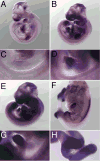
Fig. 1
Fig. 1. Wnt5a is expressed during midgut elongation
Wnt5a mRNA was detected by whole mount in situ hybridization using an antisense probe in (A,C) E9.5, (B,D) E10.0, (E,G) (E10.5) and (F,H) E12.5 embryos. (C–D) and (G–H) are higher magnification pictures of the outlined areas in (A–B) and (E–F), respectively, but they are not necessarily of the same embryos. (A,C) Expression is detected in the midgut region at E9.5 and increases over time (B,D–H). As the midgut loop starts to form at E10.0 (B,D) and E10.5 (E, G), Wnt5a expression remains restricted to this region. (F,H) Wnt5a expression persists in the midgut and hindgut mesenchyme and is excluded from the cecum at E12.5. The left limbs and the head were removed in the embryo depicted in F for clarity.
Fig. 2
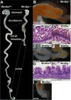
Fig. 2. Multiple defects along the gastrointestinal tract in Wnt5a mutants
All images are from E18.5 embryos. (A) The gastrointestinal tract is dramatically shortened in Wnt5a mutants compared to control littermates. The most affected segment is the small intestine. (B) The small intestine of Wnt5a mutants is marked by the presence of a bifurcation that generates a shortened gut tube parallel to the existing gut (the gut is oriented with the anterior region to the left). Hematoxylin and eosin staining of sections reveals that the small intestine is well differentiated in control (C) and Wnt5a mutants at the level of the bifurcation (D). (D) is a longitudinal section parallel to the plane of the image shown in (B). (C,G,H) are longitudinal sections. The cecum appears as a rudiment in Wnt5a mutants (F) compared to control littermates (E). Hematoxylin and eosin staining reveal the well differentiated epithelium of the large intestine in control (G) and Wnt5a mutant embryos (H). Wnt5a mutants also display an imperforate anus (I). Images in B,E,F,H were obtained using a binocular magnification lens at a 3.2x magnification. Scale bar: 100 μm.
Fig. 3
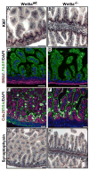
Fig. 3. Loss of Wnt5a does not alter cell differentiation in the small intestine
Immunohistochemistry on sections from Wnt5a control (A, C, E, G) and mutant (B, D, F, H) E18.5 embryos. Ki67 staining shows the proliferating intervillus region is present in control (A) and at the site of bifurcation in Wnt5a mutant (B) small intestines. The layer of smooth muscle positive for α-SMA (red) appears reduced in Wnt5a mutant small intestine (D) compared to control (C). Lfabp (green), an enterocyte marker, is appropriately expressed in Wnt5a control (C) and mutant embryos (D). Cdx2 (red), an intestinal marker, is expressed in the small intestine of control (E) and Wnt5a mutant (F) embryos. Binding of DBA (green) to the mucin-secreting cells in the small intestine of control (E) and Wnt5a mutant (F) embryos reveals the presence of goblet cells. The nerve plexus and abundant enteroendocrine cells are detected by anti-synaptophysin stainings in control (G) and Wnt5a mutant (H) tissues. Scale bar: 50 μm.
Fig. 4
Fig. 4. Defects in vitelline duct closure in Wnt5a mutant embryos
Whole mount immunofluorescence against the indicated markers shows the morphology of the closing midgut and the vitelline duct. E-cadherin (green) and Cdx2 (red) mark the epithelial cells of the gut and vitelline duct whereas DBA (red) marks its lumen in Wnt5a control (A,C,E,G) and mutant (B,D,F,H) embryos at the indicated stages. Even though the opening of the midgut progressively narrows in both control (A,C) and Wnt5a mutant embryos (B,D) at E9.5, the junction between the midgut and primitive gut is enlarged in Wnt5a mutant embryos. Whereas midgut and vitelline duct closure are completed in control embryos (E,G) by E10.0, these structures are still open in Wnt5a mutant littermates (F,H). All panels correspond to projections of multiple Z-stacks of the whole stained tissue except for G–H in which only 1μm-thick stacks were acquired to show cell morphology. Scale bar: 100 μm.
Fig. 5
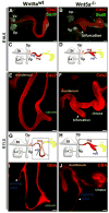
Fig. 5. Midgut elongation is abnormal in Wnt5a mutants
Whole mount immunostainings of the midgut region during its elongation in control (A,E,I) and Wnt5a mutant embryos (B,F,J) at the indicated stages. Schematics of the foregut-midgut areas at the corresponding stages are also shown for clarity (C–D,G–H). As elongation initiates at E10.5, the growing midgut forms a loop in control tissues (A) whereas a bifurcation from the main tube grows in Wnt5a mutant embryos (B) (arrowhead). The gut epithelium, stained with anti-Cdx2 (red) appears wider and shorter in Wnt5a mutants (compare A and B). The midgut loop (A,C) and the bifurcation (B,D) are formed caudal to the pancreatic buds stained with anti-Sox9 (green), between the duodenum (orange) and the cloaca (yellow) (C–D). By E11.5, the midgut loop adopts a hairpin shape and the cecum buds from the tip of the posterior portion of the hairpin in control tissues (E,G). The cecum and the posterior portion of the midgut loop (G) are shown in green and red dots, respectively. In Wnt5a mutants (F,H) the bifurcation continues to grow while the growth of the gut tube is stunted (note that E and F are the same magnification). Equivalent areas in E–G and F–H are outlined in G and H. DBA (red) marks the lumen of the gut as well as the vitelline duct which is located in the anterior portion of the hairpin loop in control embryos (I) and at the tip of the bifurcation in Wnt5a mutants (J). The vitelline duct is represented in blue in C, D, G, H. Vp: ventral pancreas, Dp: dorsal pancreas, St: stomach, Lv: liver, d: duodenum, mg: midgut, cl: cloaca, vd: vitelline duct, c: cecum. Scale bar: 100 μm.
Fig. 6
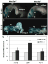
Fig. 6. Wnt/β-catenin pathway during gut elongation
Control and Wnt5a mutant foregut-midgut regions were stained with X-gal to detect TOPGAL reporter activity at E10.5 (A) and E11.5 (B). β-galactosidase activity was not detected in the midgut despite of the strong staining in the lung and forestomach in at least 2 pair of control and Wnt5a mutant embryos for each stage. Axin2, Lef1 and Tcf1 RNA quantification in E11.5 control (grey) and Wnt5a mutant (black) midgut regions (C). Bars represent the average of 5 litters normalized to both cyclophilinA and Gus expression. Error bars indicate standard deviation. Lg: lung, Dp: dorsal pancreas, St: stomach.
Fig. 7
Fig. 7. Architecture of the gut epithelium is disrupted in the absence of Wnt5a
Immunofluorescence stainings on 5 μm sections with the indicated antibodies. The gut epithelium appears mainly as a monolayer in control and Wnt5a mutant embryos stained with anti E-cadherin (green) and Cdx2 (red) antibodies at E9.5 (A–B). Staining against E-cadherin (green) shows that the elongating gut tube displays a thickened epithelium in Wnt5a mutants (C–D) at E11.5. These images show that the gut epithelium is formed by a columnar monolayer in control tissues (C). In contrast, the epithelium is multi-layered and the columnar cell morphology is lost in Wnt5a mutants (D). The epithelial architecture recovers by the onset of intestinal differentiation and villi formation by E15.5 (E–F). Quantification of cell proliferation in 4 pairs of Wnt5a control and mutant embryos at E11.5 using phospho–histone H3 as a cell proliferation marker (G). The percentage of phosphorylated Histone H3 positive cells was calculated for each embryo and the values obtained for each Wnt5a mutant embryo were normalized to the values obtained for the corresponding control littermate, which was considered to be 100%. Cell proliferation is reduced to 63.05% in Wnt5a mutant embryos (G). Dividing cells (arrowheads), stained with anti-phosphorylated-Histone H3 (blue), are located at the luminal side of the epithelium (H–I). Scale bar: 10 μm. ***P<0.005 Student’s t-test.
Fig. 8
Fig. 8. Re-intercalation of post-mitotic cells is perturbed in Wnt5a mutant gut epithelium
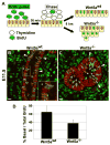
Schematics of the in vivo BrdU pulse-chase of dividing cells (A). BrdU positive post-mitotic cells are re-intercalated (arrowheads) or re-intercalating (asterisk and inset) in the Wnt5a control epithelial monolayer (B). In Wnt5a mutant midgut, few cells re-intercalate (arrowheads) whereas abundant clusters of cells are accumulated in the luminal side of the epithelium (arrows) (C). Note that the section imaged in C is at the level of the duplication, which is indicated by a ‘d’. Re-intercalated cells were quantified over several sections in 1 of the pairs of control and Wnt5a mutant embryos and found to be reduced from 64% in controls to 37% in Wnt5a mutant embryos (D). Scale bar: 10 μm.

References

    1. Ahnfelt-Rønne J, Jørgensen MC, Hald H, Madsen OD, Serup P, Hecksher-Sørensen J. An improved method for three-dimensional reconstruction of protein expression patterns in intact mouse and chicken embryos and organs. J Histochem Cytochem. 2007;55:925–30. - PubMed
    1. Bastide P, Darido C, Pannequin J, Kist R, Robine S, Marty-Double C, Bibeau F, Scherer G, Joubert D, Hollande F, Blache P, Jay P. Sox9 regulates cell proliferation and is required for Paneth cell differentiation in the intestinal epithelium. J Cell Biol. 2007;178:635–48. - PMC - PubMed
    1. Brandvold KA, Ewert DL, Kent SC, Neiman P, Ruddell A. Blocked B cell differentiation and emigration support the early growth of Myc-induced lymphomas. Oncogene. 2001;20:3226–34. - PubMed
    1. Ciruna B, Jenny A, Lee D, Mlodzik M, Schier AF. Planar cell polarity signalling couples cell division and morphogenesis during neurulation. Nature. 2006;439:220–4. - PMC - PubMed
    1. DasGupta R, Fuchs E. Multiple roles for activated LEF/TCF transcription complexes during hair follicle development and differentiation. Development. 1999;126:4557–68. - PubMed

Publication types