

Microfluidics and multielectrode array-compatible organotypic slice culture method
- PMID: 19100768
- PMCID: PMC2669279
- DOI: 10.1016/j.jneumeth.2008.11.016
Microfluidics and multielectrode array-compatible organotypic slice culture method
Abstract
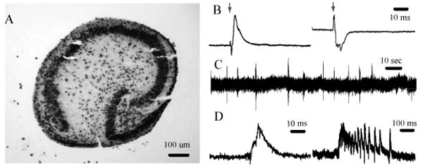
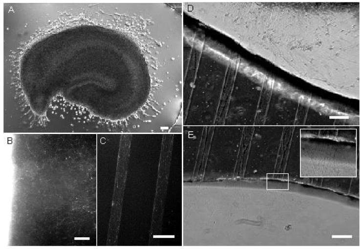
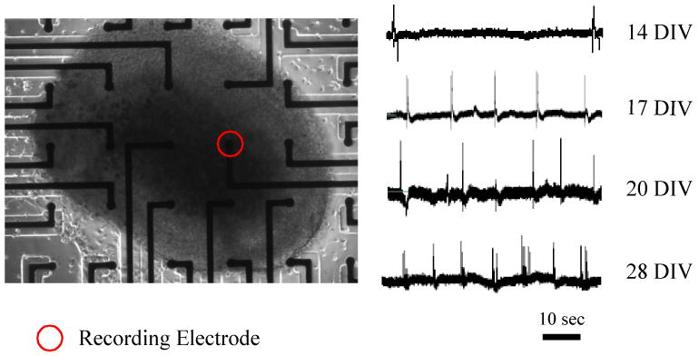
Organotypic brain slice cultures are used for a variety of molecular, electrophysiological, and imaging studies. However, the existing culture methods are difficult or expensive to apply in studies requiring long-term recordings with multielectrode arrays (MEAs). In this work, a novel method to maintain organotypic cultures of rodent hippocampus for several weeks on standard MEAs in an unmodified tissue culture incubator is described. Polydimethylsiloxane (Sylgard) mini-wells were used to stabilize organotypic cultures on glass and MEA surfaces. Hippocampus slices were successfully maintained within PDMS mini-wells for multiple weeks, with preserved pyramidal layer organization, connectivity, and activity. MEAs were used to record the development of spontaneous activity in an organotypic cultures for 4 weeks. This method is compatible with integration of microchannels into the culture substrate. Microchannels were incorporated into the mini-wells and applied to the guidance of axons originating within the slice, paving the way for studies of axonal sprouting using organotypic slices.
Figures

References
-
- Chung BG, Flanagan LA, Rhee SW, Schwartz PH, Lee AP, Monuki ES, Jeon NL. Human neural stem cell growth and differentiation in a gradient-generating microfluidic device. Lab Chip. 2005;5:401–406. - PubMed
-
- Claverol-Tinture E, Ghirardi M, Fiumara F, Rosell X, Cabestany J. Multielectrode arrays with elastomeric microstructured overlays for extracellular recordings from patterned neurons. J Neural Eng. 2005;2:L1–7. - PubMed
-
- Egert U, Schlosshauer B, Fennrich S, Nisch W, Fejtl M, Knott T, Muller T, Hammerle H. A novel organotypic long-term culture of the rat hippocampus on substrate-integrated multielectrode arrays. Brain Res Brain Res Protoc. 1998;2:229–242. - PubMed
Publication types
MeSH terms
Substances
Grants and funding
LinkOut - more resources
Full Text Sources
Other Literature Sources
