. 2009 Mar 1;46(5):616-23.
doi: 10.1016/j.freeradbiomed.2008.11.018. Epub 2008 Dec 6.

Supression of hemin-mediated oxidation of low-density lipoprotein and subsequent endothelial reactions by hydrogen sulfide (H(2)S)

Affiliations

Supression of hemin-mediated oxidation of low-density lipoprotein and subsequent endothelial reactions by hydrogen sulfide (H(2)S)

Viktória Jeney et al. Free Radic Biol Med. .

Abstract

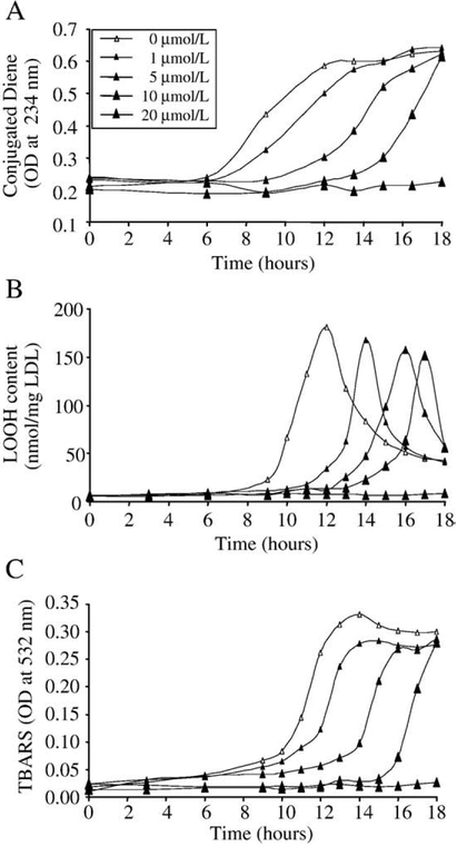
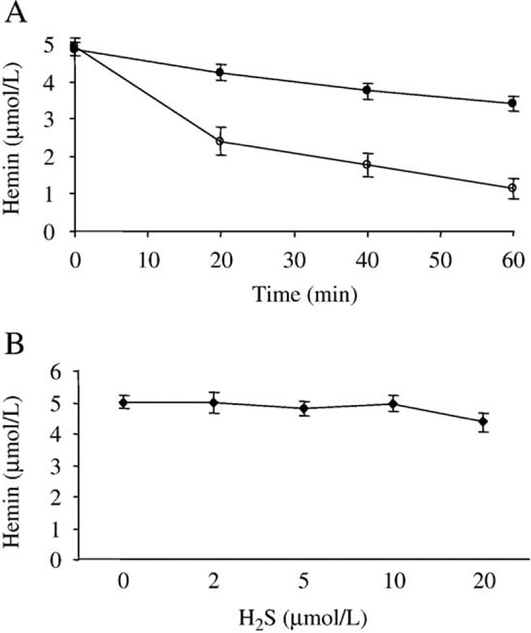
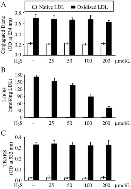
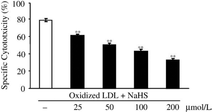
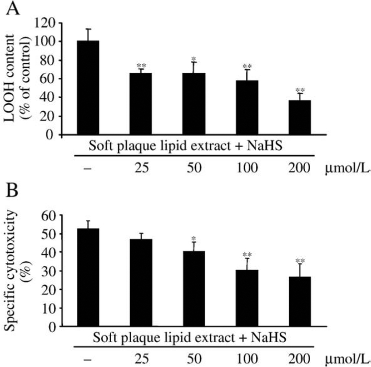
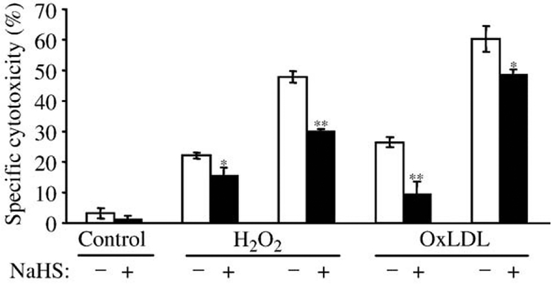
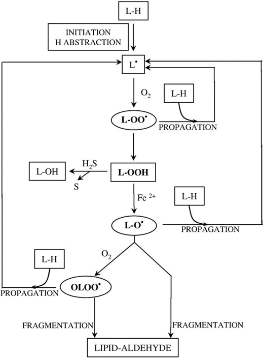
Heme-mediated oxidative modification of low-density lipoprotein (LDL) plays a crucial role in early atherogenesis. It has been shown that hydrogen sulfide (H(2)S) produced by vascular smooth muscle cells is present in plasma at a concentration of about 50 micromol/L. H(2)S is a strong reductant which can react with reactive oxygen species like superoxide anion and hydrogen peroxide. The current study investigated the effect of H(2)S on hemin-mediated oxidation of LDL and oxidized LDL (oxLDL)-induced endothelial reactions. H(2)S dose dependently delayed the accumulation of lipid peroxidation products-conjugated dienes, lipid hydroperoxides (LOOH), and thiobarbituric acid reactive substances-during hemin-mediated oxidation. Moreover, H(2)S decreased the LOOH content of both oxidized LDL and lipid extracts derived from soft atherosclerotic plaque, which was accompanied by reduced cytotoxicity. OxLDL-mediated induction of the oxidative stress responsive gene, heme oxygenase-1, was also abolished by H(2)S. Finally we have shown that H(2)S can directly protect endothelium against hydrogen peroxide and oxLDL-mediated endothelial cytotoxicity. These results demonstrate novel functions of H(2)S in preventing hemin-mediated oxidative modification of LDL, and consequent deleterious effects, suggesting a possible antiatherogenic action of H(2)S.


Figures

Fig. 1.
Fig. 1.
H2S inhibits formation of lipid peroxidation products during hemin-mediated oxidation of LDL in a dose-dependent manner. LDL (200 mg/mL) was incubated with hemin (5 mol/L) alone (empty triangle) or in the presence of H2S at a concentration of 1, 5, 10, or 20 mol/L (closed triangles) at 37°C. Lipid peroxidation was monitored by measuring the formation of conjugated dienes (A), LOOH (B), and TBARs (C) for 18 h. Figure shows a representative of three separate experiments.
Fig. 2.
Fig. 2.
Time and dose dependency of hemin scavenging by H2S. (A) Hemin (5 mmol/L) was incubated with H2S (100 μmol/L) in the absence (open circles) or in the presence of LDL (200 μg/ml) (closed circles) for the indicated time at 37°C, and then hemin content was determined as described under Materials and methods. (B) Hemin (5 μmol/L) was incubated with H2S (2–10 μmol/L) in the presence of LDL (200 μg/ml) for 6 h at 37°C, and then hemin content was determined. Figure shows mean of three independent experiments. Error bars denote standard deviations.
Fig. 3.
Fig. 3.
H2S dose dependently decreases LOOH content of oxidized LDL. LDL (200 mg/ml) was oxidized with hemin (5 μmol/L) for 12 h at 37°C, and then treated with H2S at the indicated concentrations for 30 min at 37°C. Following treatment levels of conjugated dienes (A), LOOH (B), and TBARs (C) were measured. Data are derived from five separate experiments. Error bars denote standard deviations. Statistical significance is indicated by one (Pb0.05) or two (Pb0.01) asterisks.
Fig. 4.
Fig. 4.
H2S dose dependently abolishes cytotoxic effects of oxidized LDL on HUVECs. LDL (200 μg/ml) was oxidized with hemin (5 μmol/L) for 12 h at 37°C, and then treated with H2S at the indicated concentrations for 30 min at 37°C. HUVEC grown on a 96-well plate were washed twice with HBSS and then challenged by the LDL samples for 4 h. MTT assay was performed to measure cytotoxicity. Data are derived from three separate experiments performed in triplicates. Error bars denote standard deviations. Statistical significance is indicated by one (Pb0.05) or two (Pb0.01) asterisks.
Fig. 5.
Fig. 5.
Heme oxygenase-1 induction mediated by oxLDL is diminished by H2S in a dose-responsive manner. Oxidized or native LDL (200 μg/ml) was pretreated with H2S (25, 50, 100, or 200 μmol/L) at 37°C for 30 min, and then diluted 4 times with HBSS. (A) For HO-1 mRNA detection confluent endothelial cells were treated with LDL samples (50 μg/mL) for 1 h, followed by a 4-h incubation in complete media. HO-1 mRNA copy numbers were determined by real-time RT-PCR as described under Materials and methods and were normalized by 18S rRNA copies. Fold increase was calculated using vehicle-treated cells as control (B). To determine HO-1 protein levels HUVECs grown on 6-well plates were treated with LDL samples (50 μg/ml) for 1 h, followed by a 8-h incubation in complete media. Cells were lysed and Western blot was performed as described under Materials and methods. After detection of HO-1 membrane was striped and reprobed for GAPDH to prove equal loading. Quantification of HO-1 induction was performed using computer-assisted videodensitometry. (C) HO-1 activity measured at 8 h after exposure of HUVECs to the same LDL as above. Data for panels A and C are derived from three separate experiments. Error bars denote standard deviations. Statistical significance is indicated by one (P<0.05) or two (P<0.01) asterisks. Western zblot (B) is a representative of three separate experiments.
Fig. 6.
Fig. 6.
H2S dose dependently decreases LOOH content and cytotoxicity of oxidized lipid extract derived from soft plaque. Lipid was extracted from human atherosclerotic lesions as described under Materials and methods. Oxidized extracted lipid (1 mg lipid/ml) was oxidized with hemin (5 μmol/L) for 16 h at 37°C and then treated with H2S at the indicated concentrations for 30 min at 37°C. Following H2S treatment LOOH levels of samples were determined (A). To measure the effect of H2S on the cytotoxicity of oxidized lipid extract, HUVECs grown in 96-well plates were treated with samples generated as above for 4 h, and then MTT assay was performed (B). Data are derived from three separate experiments performed in duplicates. Error bars denote standard deviations. Statistical significance is indicated by one (Pb0.05) or two (Pb0.01) asterisks.
Fig. 7.
Fig. 7.
H2S protects HUVECs from cytotoxicity mediated by both H2O2 and oxidized LDL. HUVECs grown in 96-well plates were pretreated with H2S (50 μmol/L) at 37°C for 4 h and then challenged with hemin (5 μmol/L at 37°C for 1 h), followed by oxidative stress generated by hydrogen peroxide (100 and 200 μmol/L) or oxidized LDL (100 and 200 μg/ml). After a 4-h incubation MTT assay was performed as described under Materials and methods. Data are derived from five separate experiments performed in triplicates. Error bars denote standard deviations. Statistical significance is indicated by one (Pb0.05) or two (Pb0.01) asterisks.
Fig. 8.
Fig. 8.
Proposed mechanism of the protective effect of H2S. Lipid peroxidation starts with a H atom abstraction which leads to the formation of a lipid radical (L). Lipid radical reacts with moleacular oxygen forming lipid peroxyl radical, one of the radical species which can further propagate lipid peroxidation by reacting with an adjacent polyunsaturated fatty acid producing a new lipid radical and lipid hydroperoxide (LOOH). In the presence of transition metals, e.g., iron, LOOH can form alkoxyl radicals (LO) and epoxy-allylic peroxyl radicals (OLOO) which can propagate the reaction. As LOOH is the ultimate source of alkoxyl radicals and epoxy-allylic peroxyl radicals its decomposition by H2S to lipid alcohols (L-OH) slows down lipid peroxidation.

References

    1. Steinberg D; Parthasarathy S; Carew TE; Khoo JC; Witztum IL Beyond cholesterol: modifications of low-density lipoprotein that increase its atherogenicity. N. Engl. J. Med 320:915–924; 1989. - PubMed
    1. Steinberg D Oxidative modification of LDL and atherogenesis. Circulation 95: 1062–1071; 1997. - PubMed
    1. Bunn HF; Jandl JH Exchange of heme among hemoglobins and between hemoglobin and albumin. J. Biol. Chem 243:465–475; 1968. - PubMed
    1. Balla G; Jacob HS; Eaton JW; Belcher JD; Vercellotti GM Hemin: a possible physiological mediator of low density lipoprotein oxidation and endothelial cell injury. Arterioscler. Thromb 11:1700–1711; 1991. - PubMed
    1. Abraham NG; Lavrovsky Y; Schwartzman ML; Stoltz RA; Levere RD; Gerritsen ME; Shibahara S; Kappas A Transfection of the human heme oxygenase gene into rabbit coronary microvessel endothelial cells: protective effect against heme and hemoglobin toxicity. Proc. Natl. Acad. Sci. USA 92:6798–6802; 1995. - PMC - PubMed

Publication types

MeSH terms