. 2009 Mar-Apr;126(3-4):142-59.
doi: 10.1016/j.mod.2008.11.007. Epub 2008 Dec 7.

Requirement of Wnt/beta-catenin signaling in pronephric kidney development

Affiliations

Requirement of Wnt/beta-catenin signaling in pronephric kidney development

Jon P Lyons et al. Mech Dev. 2009 Mar-Apr.

Abstract

The pronephric kidney controls water and electrolyte balance during early fish and amphibian embryogenesis. Many Wnt signaling components have been implicated in kidney development. Specifically, in Xenopus pronephric development as well as the murine metanephroi, the secreted glycoprotein Wnt-4 has been shown to be essential for renal tubule formation. Despite the importance of Wnt signals in kidney organogenesis, little is known of the definitive downstream signaling pathway(s) that mediate their effects. Here we report that inhibition of Wnt/beta-catenin signaling within the pronephric field of Xenopus results in significant losses to kidney epithelial tubulogenesis with little or no effect on adjoining axis or somite development. We find that the requirement for Wnt/beta-catenin signaling extends throughout the pronephric primordium and is essential for the development of proximal and distal tubules of the pronephros as well as for the development of the duct and glomus. Although less pronounced than effects upon later pronephric tubule differentiation, inhibition of the Wnt/beta-catenin pathway decreased expression of early pronephric mesenchymal markers indicating it is also needed in early pronephric patterning. We find that upstream inhibition of Wnt/beta-catenin signals in zebrafish likewise reduces pronephric epithelial tubulogenesis. We also find that exogenous activation of Wnt/beta-catenin signaling within the Xenopus pronephric field results in significant tubulogenic losses. Together, we propose Wnt/beta-catenin signaling is required for pronephric tubule, duct and glomus formation in Xenopus laevis, and this requirement is conserved in zebrafish pronephric tubule formation.


Figures

FIG. 1
FIG. 1
β-Engrailed or dnXTcf3 inhibit pronephric tubule and duct development. (A) Schematic overview of β-Engrailed chimera showing replacement of native β-catenin's carboxy-terminal transactivation region with the Drosophila Engrailed repression domain. Left V2 blastomeres of eight-cell stage embryos were co-injected with various doses of β-Engrailed (versus nuclear-directed β-galactosidase as RNA loading control) and rhodamine dextran lineage tracer. Embryos were fixed at stage 37-41, analyzed for correct lineage, and processed for tubule immunostaining using the 3G8 monoclonal antibody (blue) or for duct immunostaining using the 4A6 monoclonal antibody (red). (B) β-Engrailed generates loss of 3G8 kidney tubule expression as shown in the corresponding enlarged inset. (F) β-Engrailed also generates loss of 4A6 kidney duct expression as shown in the corresponding enlarged inset (tubule=blue, duct=red). (D) β-gal injected embryos, or non-injected control sides of embryos (C, E, & G), show normal 3G8 pronephric tubule and 4A6 pronephric duct expression. (H) The Pronephric Index (PNI) scoring system was utilized to quantify reductions in 3G8 tubule expression, with scores of 2.41 and 2.74 for β-Engrailed and dnXTcf3, respectively, indicating a significant loss (greater than PNI of 2.0). Uninjected and nuclear β-gal injection controls indicated no significant loss.
FIG. 2
FIG. 2
Wnt/β-catenin signaling activity in pronephric tubules of Xenopus transgenic TOPTK-iGFP reporter tadpoles. Lateral views of anterior portion of left and right sides of a representative transgenic tadpole (fixed stage 36) treated with dexamethasone (10 μM) at stage 32 for 16 hours at 15 °C. Wnt/β-catenin signaling is observed in the eyes (E), mid-brain (MBr), hind-brain (HBr) and pronephric tubules (PT, arrows).
FIG. 3
FIG. 3
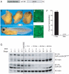
Characterization of EnR-LefΔN-GR755A, an inducible fusion construct inhibiting Wnt/β-catenin signaling. (A) EnR-LefΔN-GR755A schematic indicating the Drosophila Engrailed repressor domain (aa 1-299), mouse Lef1 DNA-binding HMG box (aa 265-391), human Glucocorticoid receptor hormone binding domain (aa 512-777), and double HA epitope tag. To abrogate potential recruitment of transcriptional coactivators, the TAF-2 motif in the GR domain was mutated (glutamine to alanine substitution at position 755). Xenopus embryos were injected with 0.5 ng EnR-LefΔN-GR755A into both animal-dorsal blastomeres at the four-cell stage and grown to stage 40 with or without dexamethasone. In the presence of dexamethasone, embryos exhibited strong ventralization phenotypes (B), while embryos not treated with dexamethasone developed normally (C). Fluorescent (FITC) immunohistochemistry of animal caps excised from stage 9/10 embryos show nuclear localization of EnR-LefΔN-GR755A in the presence of dexamethasone (D) and cytoplasmic localization in the absence of dexamethasone (E) (1.0 ng EnR-LefΔN-GR755A injected into single animal-dorsal blastomere at the 2-cell stage). (F) Embryos from panels B and C were scored using the Dorso-Anterior Index (DAI) system with embryos scoring between 0-to-4 designated positive for ventralization while embryos scoring 5 were normal. A high proportion of embryos were ventralized in the presence of dexamethasone, while none were ventralized in dexamethasone absence. (G) To assess EnR-LefΔN-GR755A protein stability following dexamethasone addition at later stages, a time course was undertaken of embryos injected with 0.5 ng EnR-LefΔN-GR755A (or control EnR-GR) into a single vegetal-ventral (V2) blastomere at the eight-cell stage. HA epitope-tag Western blotting (IB=immunoblot) shows that each chimera is stably present until addition of dexamethasone at stage 16, at which time each experiences increased metabolic turnover with dexamethasone.
FIG. 4
FIG. 4
EnR-LefΔN-GR755A inhibits pronephric epithelial tubulogenesis. (A) Injection of 0.5 ng of EnR-LefΔN-GR755A into a single V2 blastomere at the eight-cell stage and dexamethasone addition at stage 17 results in a significant or complete loss of pronephric tubule epithelium on the injected side as assessed using the 3G8 antibody. All embryos were fixed at stage 37-41. Inset shows enlargement of the 3G8-stained pronephric region. (B) The control side of the embryo shows normal 3G8 pronephric tubule staining in the presence of dexamethasone. Tubulogenesis is normal in injected or non-injected sides of non-dexamethasone treated embryos (not shown). (C) Embryo dorsal views indicate normal axis lengths with minimal axis bending. (D) Double immunostaining with 3G8 (blue) and 12/101 somite marker (red) shows EnR-Lef1ΔN-GR755A-injected embryo sides completely lack pronephric tubules while exhibiting largely normal somitogenesis (somite number unaffected and minimal effects upon segmentation). (E) Non-injected side of EnR-Lef1ΔN-GR755A-injected embryos show normal 3G8 tubule and 12/101 somite staining. (F, G) Injected and non-injected sides of EnR-GR (negative-control construct) injected embryos display normal tubule and somite development. (H) The Pronephric Index (PNI) scoring system was used to quantify the 3G8 tubule staining at stage 37/38 of EnR-LefΔN-GR755A versus control EnR-GR-injected embryos. In the plus dexamethasone condition, EnR-LefΔN-GR755A-injected embryos show a significant loss of pronephric tubulogenesis (PNI score of 2.64), while in the absence of dexamethasone no significant loss was observed. The control chimera EnR-GR (with or without dexamethasone) showed no significant loss of tubulogenesis.
FIG 5
FIG 5
EnR-LefΔN-GR755A inhibits expression of multiple pronephric glomus, epithelial tubule, and duct-specific genes. (A) Schematic showing pronephric glomus, tubule and duct segment nomenclature at stage 35. The glomus, labeled G, is shown in beige. The early and late segments within both the proximal and distal domains of tubules are shown in dark and light gray, respectively. The duct is shown in white. Injection of EnR-LefΔN-GR755A (0.5 ng) with rhodamine dextran tracer was performed into single V2 blastomeres at the eight-cell stage and dexamethasone was added at stage 16. Embryos were fixed at stages 35/36 (B-C, E-F, H-I, K-L) and expression of several pronephric marker genes analyzed by whole mount colorimetric or fluorescent in situ. Schematics represent the expression domains of four later stage pronephric markers (D, G, J, M). Shown are injected and control sides of embryos, each including insets of enlarged pronephric regions. Upon the inhibition of Wnt/β-catenin signaling, the strong late distal expression of the sodium bicarbonate cotransporter XNBC1 (slc4a4) (D) is lost on the injected (B) versus control side (C). Earlier weaker expression of this same marker is also lost (not shown). The early distal Na-K-2Cl cotransporter NKCC2 (slc12a1) (G) shows complete loss of expression on injected (E) versus control side (F). Expression of the NaK ATPase (atp1a1) in both the pronephric tubules and duct (J) is lost on the experimental side (H) as compared to the control side (I) when Wnt/β-catenin signaling is inhibited. Additionally, inhibition of Wnt/β-catenin signaling causes the normal expression of nephrin (nhps1) in the glomus (M) to be reduced in the injected side (K) as compared with the control side (L).
FIG. 6
FIG. 6
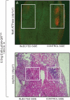
EnR-LefΔN-GR755A reduces expression of NaK ATPase and nephrin in the pronephric region. Cross sections of embryos injected with EnR-LefΔN-GR755A (left V2 blastomere at eight-cell stage), induced at stage 16, fixed at stage 35/36, and stained by in situ hybridization. (A) Fluorescent in situ hybridization of cross sections of stage 35/36 embryos shows that EnR-LefΔN-GR755A inhibits NaKATPase (atp1a1) expression within the injected side (left) versus the control side (right). (B) In situ hybridization shows that expression of nephrin (nhps1), a glomus marker, is inhibited by EnR-LefΔN-GR755A in injected (left) sides of stage 35/36 embryos as compared to control (right) sides.
FIG. 7
FIG. 7
EnR-LefΔN-GR755A generates reduced expression of pronephric mesenchyme markers. EnR-LefΔN-GR755A (0.5 ng) with rhodamine dextran tracer was injected into single V2 blastomeres at the eight-cell stage and dexamethasone was added at stage 16. Embryos were fixed at stage 23 (A&B), stage 25 (C&D), and stage 27 (E&F). The injected side of embryos shows reduced expression of pronephric mesenchyme markers XPax8 (pax8) (A), XLim1 (lhx1) (C), and vHNF1 (hnf1β) (E), shown via whole mount colorimetric or fluorescent in situ. The control side of embryos shows expected normal distribution of pronephric mesenchyme markers (B, D, F). Injected and control side images shown are from the same embryo and insets are enlargements of the pronephric region.
FIG. 8
FIG. 8
Loss of pronephric tubule tissue in zebrafish tadpoles following transgenic Dkk1 misexpression. Dorsal views of dkk1 wildtype (A) and heterozygous (B and C) transgenic embryos heat shocked several times starting at 12 hours post fertilization (5 somite stage), followed by fixation and staining at 72 hours post fertilization with the epithelial pronephric tubule and duct-specific antibody α6F. Insets show enlargements of the pronephric tubule region, with an arrow indicating weak tubule reduction (B, right side), arrowheads indicating strong reduction (C, left and right sides), and an asterisk indicating complete absence (B, left side) of α6F tubule expression. Wildtype embryos exhibit normal α6F staining of pronephric tubules (A). α6F expression in hsDkk1GFP transgenic embryos was tabulated, with 47.2% of embryos displaying a reduction or complete absence of expression (D).
FIG. 9
FIG. 9
Characterization and pronephric phenotypic analysis of LefΔN-βCTA-GR755A, an inducible fusion construct promoting Wnt/β-catenin signaling. (A) LefΔN-βCTA-GR755A schematic including the mouse Lef1 DNA-binding HMG box (aa 265-391), a double HA epitope tag, the mouse β-catenin transactivation domain, and the human Glucocorticoid receptor hormone binding domain (aa 512-777). Xenopus embryos were injected with 0.1 ng LefΔN-βCTA-GR755A into a single ventral-vegetal blastomere at the four-cell stage and grown to stage 33-35 with or without dexamethasone. In the presence of dexamethasone, embryos showed a robust level of secondary axis phenotypes (B), while embryos not treated with dexamethasone developed normally (C). Fluorescent (FITC) immunohistochemistry of animal caps excised from stage 9/10 embryos show nuclear localization of LefΔN-βCTA-GR755A in the presence of dexamethasone (D) and cytoplasmic localization in the absence of dexamethasone (E) (prior to animal cap excision, embryos were injected with 1.0 ng LefΔN-βCTA-GR755A into single animal-dorsal blastomeres at the 2-cell stage). (F) Most all embryos noted in B (presence of dexamethasone) exhibited one or more features reflecting an ectopic dorsal axis, while none of those noted in C (absence of dexamethasone) exhibited duplicate axes. (G) To assess LefΔN-βCTA-GR755A protein stability following dexamethasone addition, a time course was undertaken of embryos injected with 0.5 ng LefΔN-βCTA-GR755A into single vegetal-ventral (V2) blastomeres at the eight-cell stage. HA epitope-tag Western blotting shows the inducible chimera (like that of EnR-LefΔN-GR755A and EnR-GR) is stably present until addition of dexamethasone at stage 16, at which time each experiences increased metabolic turnover. (H) Xenopus embryos injected with 0.1 ng LefΔN-βCTA-GR755A into single vegetal-ventral (V2) blastomeres at the eight-cell stage (+dexamethasone at stage 16) showed decreased expression of an early pronephric tubule epithelial marker hnf1β (in situ at stage 26), and variable decreased expression of a more mature tubule epithelial marker 3G8 (immunostaining at stage 35). The left panels show whole tadpoles along with enlargements of observed phenotypes on injected sides, while right panels depict uninjected/control sides with corresponding enlargements.

Similar articles

Cited by

References

    1. Aulehla A, Herrmann BG. Segmentation in vertebrates: clock and gradient finally joined. Genes Dev. 2004;18:2060–7. - PubMed
    1. Benzing T, Simons M, Walz G. Wnt signaling in polycystic kidney disease. J Am Soc Nephrol. 2007;18:1389–98. - PubMed
    1. Brandli AW. Towards a molecular anatomy of the Xenopus pronephric kidney. Int J Dev Biol. 1999;43:381–95. - PubMed
    1. Brembeck FH, Rosario M, Birchmeier W. Balancing cell adhesion and Wnt signaling, the key role of beta-catenin. Curr Opin Genet Dev. 2006;16:51–9. - PubMed
    1. Bridgewater D, Cox B, Cain J, Lau A, Athaide V, Gill PS, Kuure S, Sainio K, Rosenblum ND. Canonical WNT/beta-catenin signaling is required for ureteric branching. Dev Biol. 2008;317:83–94. - PubMed

Publication types

MeSH terms

LinkOut - more resources