Review
. 2009 Feb;19(1):17-24.
doi: 10.1016/j.semcancer.2008.11.006. Epub 2008 Dec 3.

Hexokinase-2 bound to mitochondria: cancer's stygian link to the "Warburg Effect" and a pivotal target for effective therapy

Affiliations
Review

Hexokinase-2 bound to mitochondria: cancer's stygian link to the "Warburg Effect" and a pivotal target for effective therapy

Saroj P Mathupala et al. Semin Cancer Biol. 2009 Feb.

Abstract

The most common metabolic hallmark of malignant tumors, i.e., the "Warburg effect" is their propensity to metabolize glucose to lactic acid at a high rate even in the presence of oxygen. The pivotal player in this frequent cancer phenotype is mitochondrial-bound hexokinase [Bustamante E, Pedersen PL. High aerobic glycolysis of rat hepatoma cells in culture: role of mitochondrial hexokinase. Proc Natl Acad Sci USA 1977;74(9):3735-9; Bustamante E, Morris HP, Pedersen PL. Energy metabolism of tumor cells. Requirement for a form of hexokinase with a propensity for mitochondrial binding. J Biol Chem 1981;256(16):8699-704]. Now, in clinics worldwide this prominent phenotype forms the basis of one of the most common detection systems for cancer, i.e., positron emission tomography (PET). Significantly, HK-2 is the major bound hexokinase isoform expressed in cancers that exhibit a "Warburg effect". This includes most cancers that metastasize and kill their human host. By stationing itself on the outer mitochondrial membrane, HK-2 also helps immortalize cancer cells, escapes product inhibition and gains preferential access to newly synthesized ATP for phosphorylating glucose. The latter event traps this essential nutrient inside the tumor cells as glucose-6-P, some of which is funneled off to serve as carbon precursors to help promote the production of new cancer cells while much is converted to lactic acid that exits the cells. The resultant acidity likely wards off an immune response while preparing surrounding tissues for invasion. With the re-emergence and acceptance of both the "Warburg effect" as a prominent phenotype of most clinical cancers, and "metabolic targeting" as a rational therapeutic strategy, a number of laboratories are focusing on metabolite entry or exit steps. One remarkable success story [Ko YH, Smith BL, Wang Y, Pomper MG, Rini DA, Torbenson MS, et al. Advanced cancers: eradication in all cases using 3-bromopyruvate therapy to deplete ATP. Biochem Biophys Res Commun 2004;324(1):269-75] is the use of the small molecule 3-bromopyruvate (3-BP) that selectively enters and destroys the cells of large tumors in animals by targeting both HK-2 and the mitochondrial ATP synthasome. This leads to very rapid ATP depletion and tumor destruction without harm to the animals. This review focuses on the multiple roles played by HK-2 in cancer and its potential as a metabolic target for complete cancer destruction.


Figures
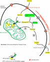
Fig. 1
Fig. 1
Metabolic channeling of glucose within a highly glycolytic tumor cell. Glucose brought across the plasma-membrane by glucose transporters is rapidly phosphorylated by HK-2 bound to VDAC located on the outer mitochondrial membrane. VDAC channels ATP generated by the ATP Synthasome complex (ATP synthase; adenine nucleotide translocator, ANT; inorganic phosphate carrier, PiC) on the inner mitochondrial membrane, facilitating direct access of ATP to VDAC-bound HK-2. To maintain the high rate of glycolytic metabolism in tumors, and their proliferation capacity, the product G-6-P rapidly distributes primarily across two key metabolic routes; (a) entry of G-6-P into the pentose–phosphate shunt for biosynthesis of nucleic-acid precursors, and (b) conversion of the G-6-P via the glycolytic pathway to pyruvic acid. Most of the pyruvic acid is reduced to lactic acid and transported out of the tumor cell via lactate transporters {B}. This promotes an unfavorable environment for the surrounding normal cells with concomitant regeneration of NAD+ within the cells to maintain glycolysis. Some pyruvate is directed to mitochondria across VDAC and via the “as-yet-uncharacterized” pyruvate transporter on the inner mitochondrial membrane {A}. This provides substrates for the tri-carboxylic acid (TCA) cycle for energy generation, as well as lipid and amino acid biosynthesis (not shown).
Fig. 2
Fig. 2
Metabolic targeting strategies against malignant tumors that rely on HK-2 facilitated glycolytic flux. The pyruvate analog 3-Br-pyruvate after entering tumor cells inhibits the function of both hexokinase-2 {1} and the ATP synthasome (ATP synthase/adenine nucleotide carrier/phosphate carrier complex) {2}. Peptide analogs of the HK-2 N-terminal, or small-molecules clotrimazole, bifanazole or methyl jasmonate, can dislodge HK-2 from VDAC {1}, depriving HK-2 of direct access to mitochondrial generated ATP and negating tolerance to G-6-P mediated product inhibition. Lactate efflux by the monocarboxylate transporters (MCTs) can be inhibited via small-molecule cinnamic acid derivatives, i.e., α-cyano-4-hydroxy cinnamic acid {3} or silenced via siRNA; tumor specific lactate dehydrogenase (LDH) isoforms can be silenced via siRNA {4}; the pyruvate analog dichloroacetate (DCA) can be administered to inhibit mitochondrial pyruvate dehydrogenase kinase (PDHK) {5}, which prevents “inactivation” of mitochondrial pyruvate dehydrogenase (PDH). The “active” PDH facilitates a high rate of influx of pyruvate into mitochondria, with concomitant up-regulation of the TCA cycle, resulting in an altered tumor-mitochondrial redox status; tumor specific pyruvate kinase (PK) can be silenced via siRNA to alter the glycolytic flux by inhibiting the formation of pyruvate {6}. Each of the above steps, which either target HK-2, the first step of the glycolytic pathway, or force the re-direction of pyruvate at the ultimate steps of the same, result in a highly disturbed metabolic status in the tumor mitochondria. This likely activates disruption of mitochondrial membrane integrity and release of cytochrome c to the cytoplasm, which in turn activates apoptotic cascades in the targeted tumor cells (MPTP, mitochondrial permeability transition pore).
Fig. 3
Fig. 3
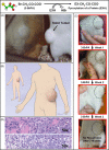
Potent anticancer effect of 3-bromopyruvate. Notably, of all the anti-cancer agents noted in the legend to Fig. 2, 3-bromopyruvate has been the most effective in completely eradicating tumors in immuno-competent animals. Significantly, this tiny agent induces a rapid loss of cellular ATP in those tumors that exhibit a robust Warburg effect, and its mode of action extends beyond apoptotic effects per se and likely involves a combination of apoptotic and necrotic events. In the figure 3-bromopyruvate completely eradicates a large rat hepatocellular carcinoma {A} that projected on a human {B} would be the size of a large “grapefruit”. Complete eradication was obtained in 3 weeks with no recurrence {C}, the animal living out a normal life thereafter. Tumor histopathology of the untreated animal {D}. In the same study 18 other animals were freed of advanced cancer. All cancer free animals lived out a normal life without return of cancer (permission granted from Elsevier to reproduce Fig. 2 from Ref. [49]).

Similar articles

Cited by

References

    1. Wilson JE. Hexokinases. Rev Physiol Biochem Pharmacol. 1995;126:65–198. - PubMed
    1. Wilson JE. An introduction to the isoenzymes of mammalian hexokinase types I-III. Biochem Soc Trans. 1997;25:103–7. - PubMed
    1. Wilson JE. Isozymes of mammalian hexokinase: structure, subcellular localization and metabolic function. J Exp Biol. 2003;206:2049–57. - PubMed
    1. Tsai HJ, Wilson JE. Functional organization of mammalian hexokinases: characterization of chimeric hexokinases constructed from the N- and C-terminal domains of the rat type I and type II isozymes. Arch Biochem Biophys. 1995;316:206–14. - PubMed
    1. Tsai HJ, Wilson JE. Functional organization of mammalian hexokinases: both N- and C-terminal halves of the rat type II isozyme possess catalytic sites. Arch Biochem Biophys. 1996;329:17–23. - PubMed

Publication types