. 2009 Mar;49(3):821-31.
doi: 10.1002/hep.22695.

Unique phenotype of hepatocellular cancers with exon-3 mutations in beta-catenin gene

Affiliations

Unique phenotype of hepatocellular cancers with exon-3 mutations in beta-catenin gene

Benjamin Cieply et al. Hepatology. 2009 Mar.

Abstract

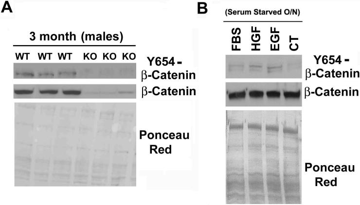
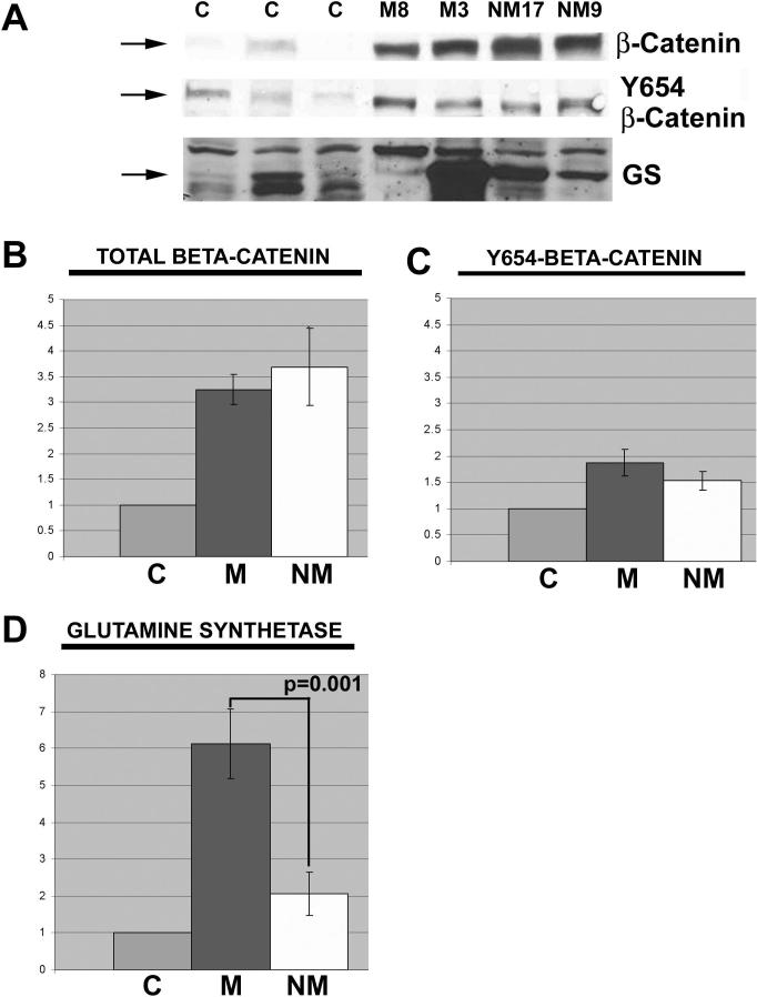
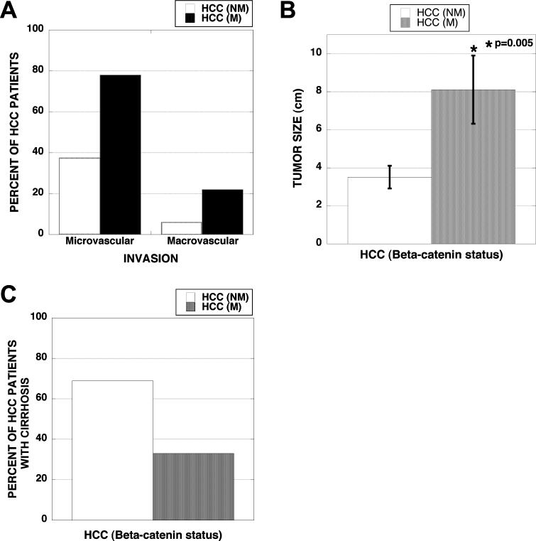
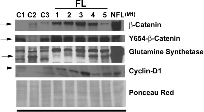
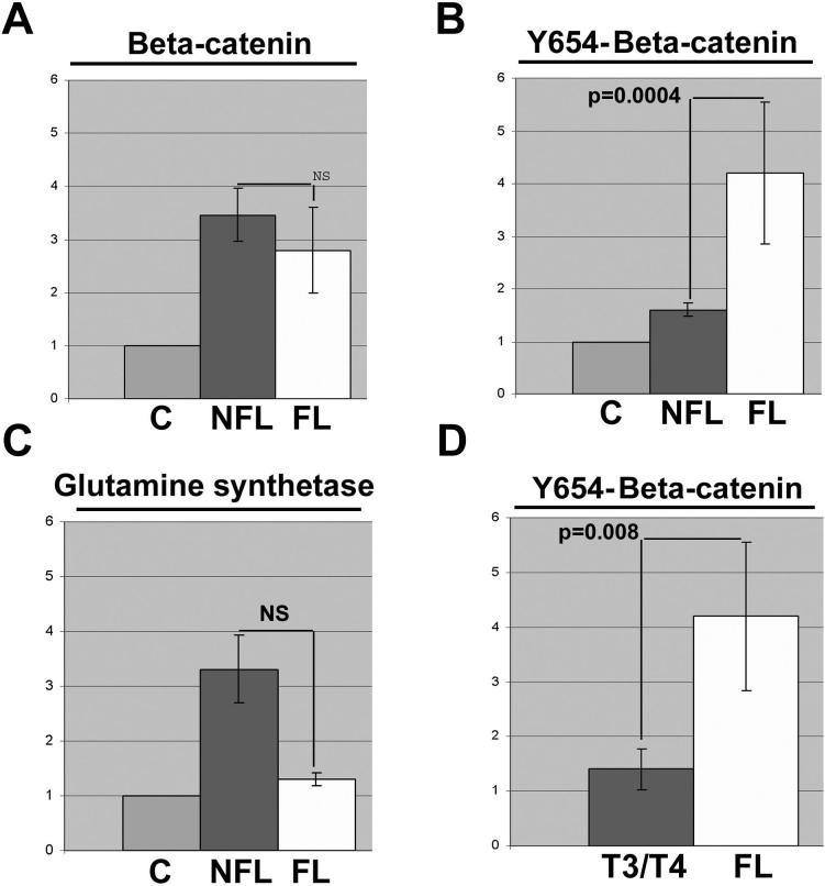
Wnt/beta-catenin signaling plays an important role in liver development and regeneration. Its aberrant activation, however, is observed in a subset of primary hepatocellular cancers (HCCs). In the current study, we compare and contrast the tumor characteristics of HCC in the presence or absence of mutations in the beta-catenin gene (CTNNB1). Frozen HCCs (n = 32), including five fibrolamellar (FL) variants, and control livers (n = 3) from Health Sciences Tissue Bank and Department of Surgery at the University of Pittsburgh Medical Center, were examined for mutations in CTNNB1, protein levels of beta-catenin, tyrosine-654-phosphorylated-beta-catenin (Y654-beta-catenin), and glutamine synthetase (GS). Missense mutations in the exon-3 of CTNNB1were identified in 9/32 HCCs. Total beta-catenin levels were higher than controls in most tumors; however, GS was exclusively increased in HCCs with mutations. Phenotypically, greater percentages of mutated HCCs showed macrovascular and microvascular invasion. Also, the tumor size was greater than double in mutated HCCs. High levels of total beta-catenin protein were observed in multinodular tumors independent of beta-catenin mutations. In addition, significant cases with mutations showed absence of cirrhosis. Finally, the highest levels of Y654-beta-catenin were exclusively observed in fibrolamellar (FL)-HCC cases.

Conclusion: Thus, HCCs that harbor missense mutations in exon-3 of CTNNB1 exhibit, histologically, a more aggressive phenotype. Also, CTNNB1 mutations might lead to HCC in the absence of cirrhosis. Finally, FL-HCC cases display a unique up-regulation of tyrosine-phosphorylated-beta-catenin, suggesting robust receptor tyrosine kinase signaling in this tumor type.


Figures

Figure 1
Figure 1. Validation of custom phospho-Y654-β-catenin antibody
(A) Total protein from wild type and β-catenin knock out mice were analyzed by western blot with the Y654-β-catenin antibody. (B) Total protein from primary rat hepatocytes treated with FBS, HGF, EGF or no serum was analyzed by western blot for Y654-β-catenin and total β-catenin. Ponceau Red staining verified equal loading of protein in both instances.
Figure 2
Figure 2. Protein quantities relative to control in tumors with and without β-catenin gene mutations
(A) Representative western blots for total β-catenin, Y654-β-catenin, and GS in HCCs with or without mutations in exon-3 of CTNNB1 as compared to three normal livers. (B) Quantitative analysis of western blots shows a significant increase (p<0.001) in total β-catenin in HCC cases with CTNNB1 mutations (M, n=9), and HCC cases without mutations (NM, n=16) over controls (C, n=3). Data for the bar graph is normalized to control. The difference of total β-catenin between CTNNB1 mutation and no mutation HCCs was insignificant (C) Quantitative analysis of Y654-β-catenin in the same data set shows insignificant differences. (D) Similar quantitative analysis displays a significant increase in GS only in HCC cases with CTNNB1 mutations. Each graph includes standard error bars.
Figure 3
Figure 3. Vascular invasion, tumor size and cirrhosis in HCC with and without CTNNB1 mutations
(A) Around 80% of HCC cases with β-catenin-gene mutations (n=9) show microvascular and 20% display macrovascular invasion. Less than 40% of non-mutated HCCs (n=16) show microvascular whereas around 6% displayed macrovascular involvement. (B) Average diameter (cm) of the tumor (largest tumor in case with multiple nodules) was significantly greater in HCC cases with β-catenin gene mutations as compared to HCCs with wild type β-catenin gene (p=0.005). (C) Only 30% of HCC cases with missense CTNNB1 mutations exhibited concomitant cirrhosis whereas more than 70% HCC cases without β-catenin mutations displayed full-blown cirrhosis.
Figure 4
Figure 4. Protein quantities relative to control in tumors with HCC with multiple nodules compared to solitary HCC tumors
(A) β-Catenin, Y654-β-catenin and GS densitometry in all cases with either multiple HCC nodules (n=9) or solitary tumors (n=15), normalized to controls. (B) β-Catenin, Y654-β-catenin, and GS densitometry in non-β-catenin-mutated cases with either multiple HCC nodules (n=6) or solitary HCC (n=9). All graphs include standard error bars and p values wherever significant differences were observed. NS-not significant.
Figure 5
Figure 5. Mutations in β-catenin gene lead to histologically more aggressive tumor phenotype than non-mutated group
All six tumors in β-catenin-gene mutated group of HCCs with tumor size greater than 5 cm (horizontal line) showed microvascular (+) and/or macrovascular (Η) invasion whereas none of the three tumors in non-mutated group that were greater than 5 cm showed any evidence of micro- or macro-vascular invasion. M-1 in the mutated group showed coexisting microvascular invasion and multiple tumor nodules (λ). Two patients in non-mutated group (NM-4 and NM-9) also showed multinodular HCC along with evidence of microvascular invasion. Finally, only 1 patient in the non-mutated group had macrovascular involvement. Thus overall, a greater number of HCC with CTNNB1 mutations stratified into histologically aggressive phenotype (Θ) than the non-mutated HCC group.
Figure 6
Figure 6. Protein analysis of FL-HCC cases for β-catenin signaling
Western blot analysis done utilizing 25μg protein was examined for total β-catenin and cyclin-D1, and 100μg protein for Y654-β-catenin, and GS. Ponceau red staining was performed to verify equal loading. C (1-3) are three normal control livers; FL (1-5) are FL-HCC cases; and NFL (M1) is a non-fibrolamellar HCC case M-1 from Table 1, which harbored exon-3 mutation in CTNNB1.
Figure 7
Figure 7. Enhanced tyrosine-phosphorylated-β-catenin levels in FL-HCC cases
(A) Total β-catenin, (B) Y654-β-catenin, and (C) GS densitometry of fibrolamellar HCCs (FL, n=5) and all other non-fibrolamellar HCC for which tissue was available for protein analysis (NFL, n=24), relative to three control livers, identifies a unique and dramatic increase in Y654-β-catenin in FL-HCC livers. (D) Densitometry of Y654-β-catenin western blots comparing non-fibrolamellar advanced stage tumors (T3/T4) to FL-HCCs. Graphs include standard error bars and significant p values; NS indicates not statistically significant difference.

References

    1. El-Serag HB. Hepatocellular carcinoma: recent trends in the United States. Gastroenterology. 2004;127:S27–34. - PubMed
    1. Parkin DM, Bray F, Ferlay J, Pisani P. Global cancer statistics, 2002. CA Cancer J Clin. 2005;55:74–108. - PubMed
    1. Bergsland EK, Venook AP. Hepatocellular carcinoma. Curr Opin Oncol. 2000;12:357–361. - PubMed
    1. McKillop IH, Moran DM, Jin X, Koniaris LG. Molecular pathogenesis of hepatocellular carcinoma. J Surg Res. 2006;136:125–135. - PubMed
    1. Thompson MD, Monga SP. WNT/beta-catenin signaling in liver health and disease. Hepatology. 2007;45:1298–1305. - PubMed

Publication types

MeSH terms

Substances