

Is intracranial atherosclerosis an independent risk factor for cerebral atrophy? A retrospective evaluation
- PMID: 19102733
- PMCID: PMC2630977
- DOI: 10.1186/1471-2377-8-51
Is intracranial atherosclerosis an independent risk factor for cerebral atrophy? A retrospective evaluation
Abstract
Background: Our purpose was to study the association between the intracranial atherosclerosis as measured by cavernous carotid artery calcification (ICAC) observed on head CT and atrophic changes of supra-tentorial brain demonstrated by MRI.
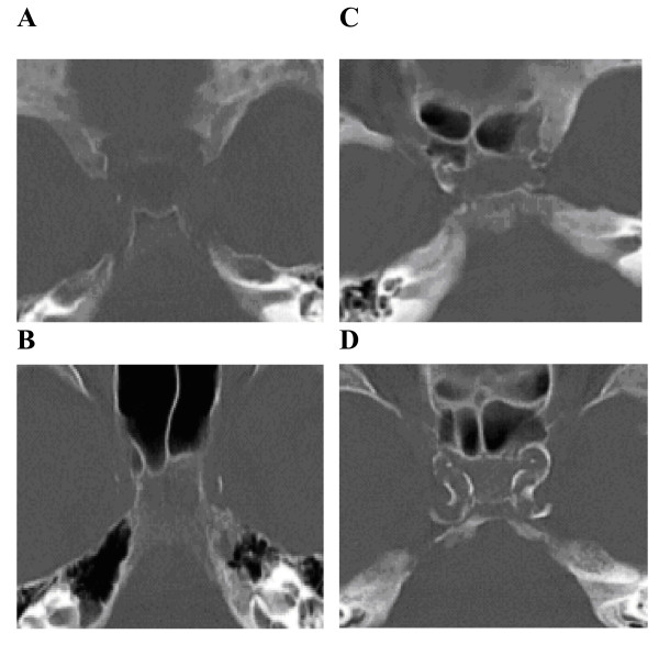
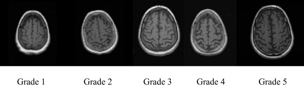
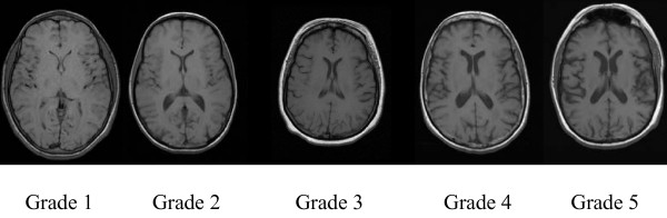
Methods: Institutional review board approval was obtained for this retrospective study incorporating 65 consecutive patients presenting acutely who had both head CT and MRI. Arterial calcifications of the intracranial cavernous carotids (ICAC) were assigned a number (1 to 4) in the bone window images from CT scans. These 4 groups were then combined into high (grades 3 and 4) and low calcium (grades 1 and 2) subgroups. Brain MRI was independently evaluated to identify cortical and central atrophy. Demographics and cardiovascular risk factors were evaluated in subjects with high and low ICAC. Relationship between CT demonstrated ICAC and brain atrophy patterns were evaluated both without and with adjustment for cerebral ischemic scores and cardiovascular risk factors.
Results: Forty-six of the 65 (71%) patients had high ICAC on head CT. Subjects with high ICAC were older, and had higher prevalence of hypertension, diabetes, coronary artery disease (CAD), atrial fibrillation and history of previous stroke (CVA) compared to those with low ICAC. Age demonstrated strong correlation with both supratentorial atrophy patterns. There was no correlation between ICAC and cortical atrophy. There was correlation however between central atrophy and ICAC. This persisted even after adjustment for age.
Conclusion: Age is the most important determinant of atrophic cerebral changes. However, high ICAC demonstrated age independent association with central atrophy.
Figures
Similar articles
-
Intracranial carotid artery calcification on head CT and its association with ischemic changes on brain MRI in patients presenting with stroke-like symptoms: retrospective analysis.Neuroradiology. 2007 Jan;49(1):27-33. doi: 10.1007/s00234-006-0159-z. Epub 2006 Nov 7. Neuroradiology. 2007. PMID: 17089112
-
Intracranial arteriosclerosis is related to cerebral small vessel disease: a prospective cohort study.Neurobiol Aging. 2021 Sep;105:16-24. doi: 10.1016/j.neurobiolaging.2021.04.005. Epub 2021 Apr 22. Neurobiol Aging. 2021. PMID: 34004492
-
Clinical and imaging features associated with intracranial internal carotid artery calcifications in patients with ischemic stroke.Neuroradiology. 2015 May;57(5):501-6. doi: 10.1007/s00234-015-1494-8. Epub 2015 Jan 30. Neuroradiology. 2015. PMID: 25633540
-
Intracranial Carotid Arteriosclerosis Mediates the Association Between Blood Pressure and Cerebral Small Vessel Disease.Hypertension. 2023 Mar;80(3):618-628. doi: 10.1161/HYPERTENSIONAHA.122.20434. Epub 2022 Dec 2. Hypertension. 2023. PMID: 36458543 Free PMC article.
-
Carotid artery constriction in autoimmune hypophysitis: three case reports and literature review.Endocr Connect. 2025 May 28;14(6):e250120. doi: 10.1530/EC-25-0120. Print 2025 Jun 1. Endocr Connect. 2025. PMID: 40337856 Free PMC article. Review.
Cited by
-
Quantification of intracranial internal carotid artery calcification on brain unenhanced CT: evaluation of its feasibility and assessment of the reliability of visual grading scales.Eur Radiol. 2013 Jan;23(1):20-7. doi: 10.1007/s00330-012-2586-z. Epub 2012 Jul 27. Eur Radiol. 2013. PMID: 22836162
-
Analysis of the tortuosity of the internal carotid artery in the cavernous sinus.Childs Nerv Syst. 2015 Jun;31(6):941-4. doi: 10.1007/s00381-015-2674-x. Epub 2015 Mar 7. Childs Nerv Syst. 2015. PMID: 25749877
-
Association of carotid and intracranial stenosis with Alzheimer's disease biomarkers.Alzheimers Res Ther. 2020 Sep 10;12(1):106. doi: 10.1186/s13195-020-00675-6. Alzheimers Res Ther. 2020. PMID: 32912336 Free PMC article.
-
Helicobacter pylori Infection Acts as an Independent Risk Factor for Intracranial Atherosclerosis in Women Less Than 60 Years Old.Front Cardiovasc Med. 2022 Jan 11;8:819315. doi: 10.3389/fcvm.2021.819315. eCollection 2021. Front Cardiovasc Med. 2022. PMID: 35087887 Free PMC article.
-
The Carotid Siphon as a Pulsatility Modulator for Brain Protection: Role of Arterial Calcification Formation.J Pers Med. 2025 Aug 4;15(8):356. doi: 10.3390/jpm15080356. J Pers Med. 2025. PMID: 40863418 Free PMC article. Review.
References
-
- Brinkman SD, Sarwar M, Levin HS, Morris HH. Quantitative indexes of computed tomography in dementia and normal aging. Radiology. 1981;138:89–92. - PubMed
-
- Ylikoski A, Erkinjuntti T, Raininko R, Sarna S, Sulkava R, Tilvis R. White matter hyperintensities on MRI in the neurologically nondiseased elderly. Analysis of cohorts of consecutive subjects aged 55 to 85 years living at home. Stroke. 1995;26:1171–7. - PubMed
MeSH terms
LinkOut - more resources
Full Text Sources
Miscellaneous
