

Expression of centromere protein F (CENP-F) associated with higher FDG uptake on PET/CT, detected by cDNA microarray, predicts high-risk patients with primary breast cancer
- PMID: 19102762
- PMCID: PMC2631591
- DOI: 10.1186/1471-2407-8-384
Expression of centromere protein F (CENP-F) associated with higher FDG uptake on PET/CT, detected by cDNA microarray, predicts high-risk patients with primary breast cancer
Abstract
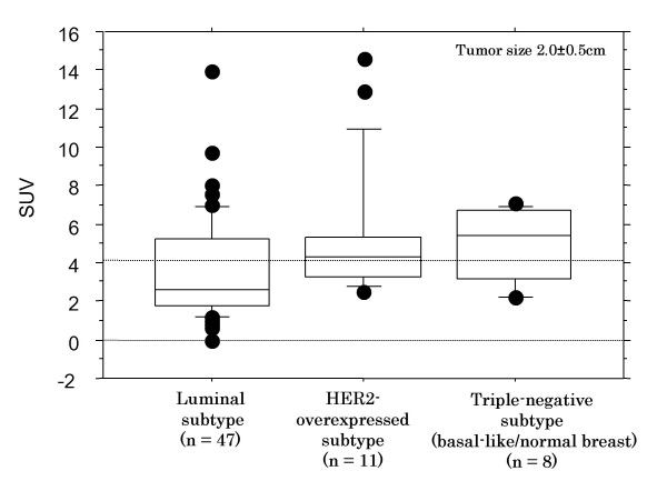
Background: Higher standardized uptake value (SUV) detected by 18F-fluorodeoxyglucose positron emission tomography/computed tomography (FDG PET/CT) correlates with proliferation of primary breast cancer. The purpose of this study is to identify specific molecules upregulated in primary breast cancers with a high SUV and to examine their clinical significance.
Methods: We compared mRNA expression profiles between 14 tumors with low SUVs and 24 tumors with high SUVs by cDNA microarray. We identified centromere protein F (CENP-F) and CDC6 were upregulated in tumors with high SUVs. RT-PCR and immunohistochemical analyses were performed to validate these data. Clinical implication of CENP-F and CDC6 was examined for 253 archival breast cancers by the tissue microarray.
Results: The relative ratios of CENP-F and CDC6 expression levels to beta-actin were confirmed to be significantly higher in high SUV tumors than in low SUV tumors (p = 0.027 and 0.025, respectively) by RT-PCR. In immunohistochemical analysis of 47 node-negative tumors, the CENP-F expression was significantly higher in the high SUV tumors (74%) than the low SUV tumors (45%) (p = 0.04), but membranous and cytoplasmic CDC6 expressions did not significantly differ between both groups (p = 0.9 each). By the tissue microarray, CENP-F (HR = 2.94) as well as tumor size (HR = 4.49), nodal positivity (HR = 4.1), and Ki67 (HR = 2.05) showed independent impact on the patients' prognosis.
Conclusion: High CENP-F expression, correlated with high SUV, was the prognostic indicators of primary breast cancer. Tumoral SUV levels may serve as a pretherapeutic indicator of aggressiveness of breast cancer.
Figures


Similar articles
-
Clinicopathological and prognostic relevance of uptake level using 18F-fluorodeoxyglucose positron emission tomography/computed tomography fusion imaging (18F-FDG PET/CT) in primary breast cancer.Jpn J Clin Oncol. 2008 Apr;38(4):250-8. doi: 10.1093/jjco/hyn019. Jpn J Clin Oncol. 2008. PMID: 18407934
-
Prognostic significance and therapeutic implications of centromere protein F expression in human nasopharyngeal carcinoma.Mol Cancer. 2010 Sep 9;9:237. doi: 10.1186/1476-4598-9-237. Mol Cancer. 2010. PMID: 20828406 Free PMC article.
-
CENP-F expression is associated with poor prognosis and chromosomal instability in patients with primary breast cancer.Int J Cancer. 2007 Apr 1;120(7):1434-43. doi: 10.1002/ijc.22413. Int J Cancer. 2007. PMID: 17205517 Free PMC article.
-
The prognostic value of SUVmax measuring on primary lesion and ALN by 18F-FDG PET or PET/CT in patients with breast cancer.Eur J Radiol. 2018 Aug;105:1-7. doi: 10.1016/j.ejrad.2018.05.014. Epub 2018 May 17. Eur J Radiol. 2018. PMID: 30017264 Review.
-
Centromere Protein F in Tumor Biology: Cancer's Achilles Heel.Cancer Med. 2025 May;14(10):e70949. doi: 10.1002/cam4.70949. Cancer Med. 2025. PMID: 40387105 Free PMC article. Review.
Cited by
-
Newly identified breast luminal progenitor and gestational stem cell populations likely give rise to HER2-overexpressing and basal-like breast cancers.Discov Oncol. 2022 May 28;13(1):38. doi: 10.1007/s12672-022-00500-6. Discov Oncol. 2022. PMID: 35633393 Free PMC article.
-
Imaging primary prostate cancer with 11C-Choline PET/CT: relation to tumour stage, Gleason score and biomarkers of biologic aggressiveness.Radiol Oncol. 2012 Sep;46(3):179-88. doi: 10.2478/v10019-012-0034-y. Epub 2012 Jun 19. Radiol Oncol. 2012. PMID: 23077456 Free PMC article.
-
Small activating RNA restores the activity of the tumor suppressor HIC-1 on breast cancer.PLoS One. 2014 Jan 28;9(1):e86486. doi: 10.1371/journal.pone.0086486. eCollection 2014. PLoS One. 2014. PMID: 24489730 Free PMC article.
-
Quantitative assessment of diffusion-weighted MR imaging in patients with primary rectal cancer: correlation with FDG-PET/CT.Mol Imaging Biol. 2011 Oct;13(5):1020-8. doi: 10.1007/s11307-010-0433-7. Epub 2010 Sep 25. Mol Imaging Biol. 2011. PMID: 20872077 Free PMC article.
-
Correlation between centromere protein-F autoantibodies and cancer analyzed by enzyme-linked immunosorbent assay.Mol Cancer. 2013 Aug 26;12(1):95. doi: 10.1186/1476-4598-12-95. Mol Cancer. 2013. PMID: 23978088 Free PMC article.
References
-
- Charlson ME, Feinstein AR. Rate of disease progression in breast cancer: a clinical estimate of prognosis within nodal and anatomic stages. J Natl Cancer Inst. 1984;72:225–231. - PubMed
Publication types
MeSH terms
Substances
LinkOut - more resources
Full Text Sources
