. 2008 Dec 23:5:117.
doi: 10.1186/1742-4690-5-117.

Human cellular microRNA hsa-miR-29a interferes with viral nef protein expression and HIV-1 replication

Affiliations

Human cellular microRNA hsa-miR-29a interferes with viral nef protein expression and HIV-1 replication

Jasmine K Ahluwalia et al. Retrovirology. .

Abstract

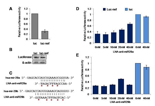
Background: Cellular miRNAs play an important role in the regulation of gene expression in eukaryotes. Recently, miRNAs have also been shown to be able to target and inhibit viral gene expression. Computational predictions revealed earlier that the HIV-1 genome includes regions that may be potentially targeted by human miRNAs. Here we report the functionality of predicted miR-29a target site in the HIV-1 nef gene.

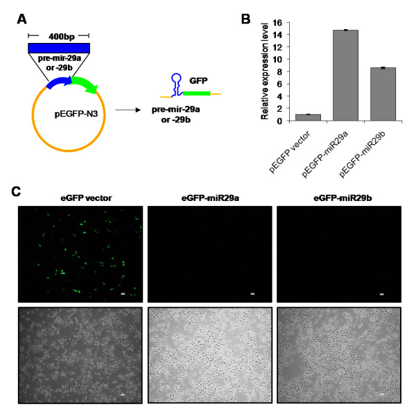
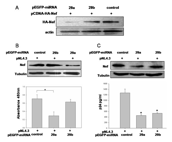
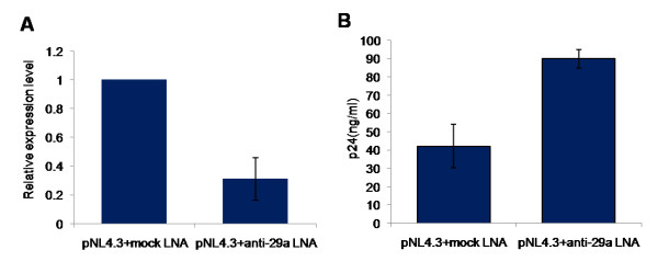
Results: We find that the human miRNAs hsa-miR-29a and 29b are expressed in human peripheral blood mononuclear cells. Expression of a luciferase reporter bearing the nef miR-29a target site was decreased compared to the luciferase construct without the target site. Locked nucleic acid modified anti-miRNAs targeted against hsa-miR-29a and 29b specifically reversed the inhibitory effect mediated by cellular miRNAs on the target site. Ectopic expression of the miRNA results in repression of the target Nef protein and reduction of virus levels.

Conclusion: Our results show that the cellular miRNA hsa-miR29a downregulates the expression of Nef protein and interferes with HIV-1 replication.


Figures

Figure 1
Figure 1
Detection of hsa-miR-29a, b, c in different cell types. (A) Schematic representation of the HIV-1 genome. Potential target sites complementary to hsa-miR-29a and b are marked with an arrow. (B) Schematic representation of the method of detection and the oligonucleotides used to capture the miRNA(s). The oligonucleotides (blue) prime sequence specific extension (green) of each miRNA due to differences at the 3' end of the oligonucleotide-miRNA hybrid. The extension product is radiolabeled by the introduction of alpha-P32-dCTP into the product at the positions underlined. The T tail of varying lengths at the 5' end is used to improve the resolution of the products. (C) Expression of hsa-miR-29a, b, c in PBMC (upper panel), HeLa (middle panel) and HEK293T (lower panel) cells. HEK293T cells show much lower expression level of hsa-miR-29a and b than PBMCs or HeLa cells. (D) U6 RNA was detected using a similar approach to establish equal input of RNA. Product sizes (nucleotides, nt) including length of T tails are indicated on the right. M: radiolabeled marker.
Figure 2
Figure 2
Nef target region downregulated reporter (luciferase) activity at a post-transcriptional level (A and B) while LNA modified anti-miRNA restored reporter activity (C, D and E). (A) A construct containing the nef target region cloned into the MCS (3'UTR) of pMIR-REPORT™ Luciferase vector was co-transfected into HeLa cells along with pMIR-REPORT™ β-gal vector. After 24 h, luciferase activity was measured and normalized to beta-galactosidase levels. Relative Luciferase activity was calculated with respect to cells transfected with pMIR-REPORT™ vector alone. Data represent mean ± SEM of three independent experiments performed, each in triplicates. (B) Post-transcriptional regulation of Nef. Northern blot using luciferase probe to show transcript levels of luciferase in luc and luc-nef transfected HeLa cells. β-actin was used as a loading control (lower panel). (C) Design of LNA-modified anti-miRNA molecules for hsa-miR-29a and 29b. Red asterisks indicate positions of LNA-modification in the backbone of the anti-miRNA molecule. (D and E) LNA-modified anti-miRNA against hsa-miR-29a (D) and hsa-miR-29b (E) restored reporter activity from the luc-nef transfected cells. Varying concentrations (0 nM–40 nM) of LNA-modified anti-miRNA molecules were co-transfected with luc-nef clone and control pMIR-REPORT™ β-gal vector into HeLa cells. Luciferase activity was measured after 24 hours and normalized to beta-galactosidase levels. Luciferase activity relative to vector (luc) was plotted. Mean ± SEM from three replicate experiments are shown. LNA, locked nucleic acid; luc, pMIR-REPORT™ vector; and luc-nef, nef target cloned in 3'UTR of pMIR-REPORT™.
Figure 3
Figure 3
pEGFP-miRNA construct expressing hsa-mir-29a and 29b. (A) Diagrammatic representation of pEGFP-N3 vector containing pre-miR29a and 29b. (B) Expression of hsa-mir-29a and 29b confirmed by quantitative real-time PCR. Data show the expression levels of mir-29a and 29b from pEGFP-miR29a and 29b constructs compared to pEGFP-N3 vector. The data represent mean ± S.D. from two independent experiments in triplicates and have been normalized to mir-92 levels. (C) GFP expression from pEGFP-miRNA transfected HEK293T cells. Upper panel represents GFP (+)ve cells, while the lower panel shows total number of cells transfected with pEGFP-N3 (left), pEGFP-miR29a (middle) and pEGFP-miR29b (right), under the 10× objective of a NIKON florescent microscope.
Figure 4
Figure 4
hsa-mir-29a and b inhibited Nef expression and HIV-1 replication. (A) Nef expression was inhibited by hsa-miR-29a and 29b. HEK293T cells were co-transfected with miRNA clones or control vector along with pCDNA-HA-Nef using calcium phosphate precipitation. After 36 hours of transfection, cells were lysed and expression of Nef was analyzed by immunoblotting using HA antibody. Immunoreactive actin bands were used as loading control. (B and C) hsa-miR-29a and hsa-miR-29b miRNA clones inhibited virus production in HEK293T (B) and Jurkat cells (C). Cells were co-transfected with miRNA clones or control vector along with HIV-1 molecular clone pNL4.3 (Materials and Methods). Cells were lysed post-transfection and expression of Nef was analyzed by immunoblotting using Nef antibody (upper panels); culture supernatant was used for p24 antigen ELISA (lower panels). Asterisks in (B) represent significant p-value of 0.016 for inhibition mediated by 29a compared to control. The difference observed with 29b is not significant. Asterisks in (C) represent significant p-value of 0.014 and 0.016 for inhibition by 29a and 29b respectively, as compared to control vector.
Figure 5
Figure 5
Cellular hsa-mir-29a inhibits HIV-1 replication. (A) LNA-modified anti-29a affected the levels of endogenous hsa-mir-29a. LNA-modified anti-29a was co-transfected with pNL4.3 vector. Real time TaqMan microRNA quantification was performed for miR29a. hsa-mir-92 was used as an endogenous control. (B) LNA-modified anti-29a increased HIV-1 replication relative to mock LNA. p24 antigen ELISA was carried out as described previously. Data represent mean ± SEM from two independent experiments performed in triplicates.

Similar articles

Cited by

References

    1. Pillai RS, Bhattacharyya SN, Filipowicz W. Repression of protein synthesis by miRNAs: how many mechanisms? Trends Cell Biol. 2007;17:118–126. doi: 10.1016/j.tcb.2006.12.007. - DOI - PubMed
    1. Callis TE, Chen JF, Wang DZ. MicroRNAs in skeletal and cardiac muscle development. DNA Cell Biol. 2007;26:219–225. doi: 10.1089/dna.2006.0556. - DOI - PubMed
    1. Fabbri M, Ivan M, Cimmino A, Negrini M, Calin GA. Regulatory mechanisms of microRNAs involvement in cancer. Expert Opin Biol Ther. 2007;7:1009–1019. doi: 10.1517/14712598.7.7.1009. - DOI - PubMed
    1. Kloosterman WP, Lagendijk AK, Ketting RF, Moulton JD, Plasterk RH. Targeted inhibition of miRNA maturation with morpholinos reveals a role for miR-375 in pancreatic islet development. PLoS Biol. 2007;5:e203. doi: 10.1371/journal.pbio.0050203. - DOI - PMC - PubMed
    1. Makeyev EV, Zhang J, Carrasco MA, Maniatis T. The MicroRNA miR-124 promotes neuronal differentiation by triggering brain-specific alternative pre-mRNA splicing. Mol Cell. 2007;27:435–448. doi: 10.1016/j.molcel.2007.07.015. - DOI - PMC - PubMed

Publication types

Substances

LinkOut - more resources