. 2009 Apr;164(2):153-6.
doi: 10.1016/j.molbiopara.2008.11.011. Epub 2008 Dec 6.

Plasmodium falciparum secretory pathway: characterization of PfStx1, a plasma membrane Qa-SNARE

Affiliations

Plasmodium falciparum secretory pathway: characterization of PfStx1, a plasma membrane Qa-SNARE

Lindsay A Parish et al. Mol Biochem Parasitol. 2009 Apr.

Abstract

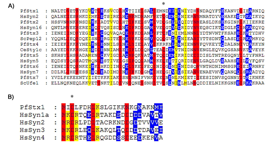
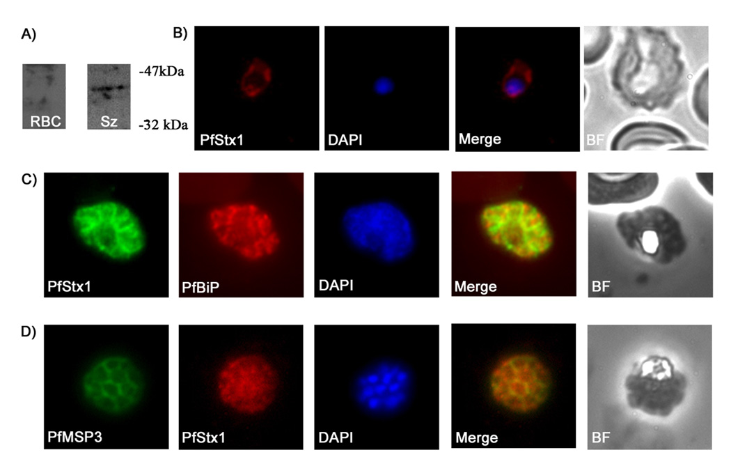
SNAREs (soluble N-ethylmaleimide-sensitive factor attachment protein receptors) play a central role in regulating and facilitating vesicular traffic in eukaryotic cells. While SNAREs have been well characterized in other eukaryotes, little is known about their role in the unique protein trafficking pathways in Plasmodium falciparum. We have identified seven Qa-SNAREs in the P. falciparum genome and confirmed the gene structure of all seven, which in one case differs from the predicted structure in the database. Based on comprehensive sequence alignments we made predictions for the intracellular locations of all seven P. falciparum Qa-SNAREs, and confirmed the predicted location for one Qa-SNARE, PfStx1, which is most closely related to other eukaryotic plasma membrane Qa-SNAREs such as syntaxin 1. This is the first identified trafficking component localized proximal to the P. falciparum plasma membrane.


Figures

Figure 1
Figure 1. Sequence alignment of PfStx proteins with other eukaryotic Qa-SNAREs
A) Sequence alignment of the SNARE domain of all seven P. falciparum Qa-SNAREs and their closest Qa-SNARE homolog from other eukaryotes. Red indicates regions where there is 80% or more similarity, yellow indicates regions where there is 60% or more similarity, and blue indicates regions where there is 40% or more similarity. The conserved Q amino acid at the core of the SNARE domain is indicated by an asterisk. B) Alignment of the N-terminus of PfStx1 with the conserved N-terminal sequence of HsSyn1a, HsSyn2, HsSyn3, HsSyn4, all of which are plasma membrane Qa-SNAREs. The first 10 amino acids of HsSyn1a interact with the SM family protein Munc18 [18], and the D3 amino acid residue (asterisk) is essential for this interaction. Residues critical for the HsSyn1a-Munc18 interaction, including D3, are conserved in PfStx1.
Figure 2
Figure 2. PfStx1 is localized proximal to the P. falciparum plasma membrane
A) Western blot of P. falciparum protein extracts run on a 12% SDS gel and probed with anti-PfStx1 antiserum. Polyclonal anti-PfStx1 antiserum was raised against a his-tagged fragment of PfStx1. A partial sequence of PfStx1 (forward primer 5’-CGGGATCCGAATTAAGTATAGAATTAAAT-3’, reverse primer 5’-CGGGAATTCTTATCTAGCTTCTATTAAATCTAC-3’) was cloned into expression plasmid pRSETA (Invitrogen) and transformed into Escherichia coli BL21-DE3 pLysS. The resulting protein was expressed as a hexa-his fusion protein and was purified from E. coli pellets by resuspending the pellets in binding buffer (8M urea, 50mM TRIS pH 8.2, 100mM NaCl and 10mM imidazole) and passing through a Hi-Trap chelating HP column (Amersham Biosciences). Columns were washed 5 times with binding buffer and purified protein was eluted with increasing amounts of imidazole ranging from 30mM to 500mM. The purified protein was then used to immunize rabbits and rats (Cocalico Biological, Inc.). Lanes: RBC – total SDS extract of uninfected erythrocytes; Sz - total SDS extract from P. falciparum 3D7 schizont stage parasites. B–D) Immunofluorescence microscopy of fixed smears of P. falciparum parasites using anti-PfStx1 antiserum. Fixed air-dried smears of P. falciparum 3D7 were fixed in a 1:1 acetone methanol solution at −20°C for 30 minutes, washed with 1X PBS and then probed with the primary antisera. Secondary antisera (Molecular Probes) labeled with a fluorophore were used for visualization on an Olympus BX60 fluorescence microscope, and images were merged with Adobe Photoshop 5.0. In trophozoite stage parasites PfStx1 (red) has a plasma membrane like staining pattern and does not appear to be exported into the erythrocyte (B). In schizont stage parasites, PfSyn1 (green) again has a peripheral location that is distinct from the ER marker PfBiP (in red) (C). PfStx1 (red) colocalizes with PfMSP3 (in green), a plasma membrane marker in schizont stage parasites (D), confirming its predicted location proximal to the plasma membrane.

Similar articles

Cited by

References

    1. Snow RW, Guerra CA, Noor AM, Myint HY, Hay SI. The global distribution of clinical episodes of Plasmodium falciparum malaria. Nature. 2005;434:214–217. - PMC - PubMed
    1. Struck NS, Herrmann S, Schmuck-Barkmann I, de Souza Dias S, Haase S, Cabrera AL, Treeck M, Bruns C, Langer C, Cowman AF, Marti M, Spielmann T, Gilberger TW. Spatial dissection of the cis- and trans-Golgi compartments in the malaria parasite Plasmodium falciparum. Mol Microbiol. 2008;67:1320–1330. - PubMed
    1. Lee MC, Moura PA, Miller EA, Fidock DA. Plasmodium falciparum Sec24 marks transitional ER that exports a model cargo via a diacidic motif. Mol Microbiol. 2008;68:1535–1546. - PMC - PubMed
    1. Yayon A, Timberg R, Friedman S, Ginsburg H. Effects of chloroquine on the feeding mechanism of the intraerythrocytic human malarial parasite Plasmodium falciparum. J Protozool. 1984;31:367–372. - PubMed
    1. Rudzinska MA, Trager W, Bray RS. Pinocytotic uptake and the digestion of hemoglobin in malaria parasites. J Protozool. 1965;12:563–576. - PubMed

Publication types

Associated data

LinkOut - more resources