

Computerized assessment of vessel morphological changes during treatment of glioblastoma multiforme: report of a case imaged serially by MRA over four years
- PMID: 19103295
- PMCID: PMC2752720
- DOI: 10.1016/j.neuroimage.2008.10.067
Computerized assessment of vessel morphological changes during treatment of glioblastoma multiforme: report of a case imaged serially by MRA over four years
Abstract
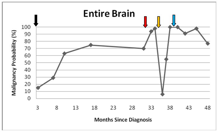
A patient with glioblastoma multiforme underwent serial computerized analysis of tumor-associated vasculature defined from magnetic resonance angiographic (MRA) scans obtained over almost a four year period. The clinical course included tumor resection with subsequent radiation therapy, a long symptom-free interval, emergence of a new malignant focus, resection of that focus, a stroke, and treatment with chemotherapy and anti-angiogenic therapy. Image analysis methods included segmentation of vessels from each MRA and statistical comparison of vessel morphology over 4 regions of interest (the initial tumor site, the second tumor site, a distant control region, and the entire brain) to the same 4 regions of interest in 50 healthy volunteers (26 females and 24 males; mean age 39 years). Results suggested that following completion of focal radiation therapy (RT) vessel shape abnormalities, if elevated at the time of RT completion, may progressively normalize for months in focal regions, that progressively severe vessel shape abnormalities can precede the emergence of a gadolinium enhancing lesion by months, that lesion resection can produce a dramatic but highly transient drop in abnormal vessel tortuosity both focally and globally, and that treatment with anti-angiogenic agents does not necessarily normalize vessel shape. Quantitative measurements of vessel morphology as defined from MRA may provide useful insights into tumor development and response to therapy.
Conflict of interest statement
Software for generic vessel segmentation has been licensed to Medtronic Corp (Minn., Minn), R2 Technologies (Alta Vista CA), Kitware (Rochester NY), and WL Gore and Associates (Flagstaff, AZ). The software has additionally been licensed for research use to multiple universities. No licensed group has been involved with the currently described work either directly or indirectly.
Figures


Similar articles
-
Fractal analysis of the susceptibility weighted imaging patterns in malignant brain tumors during antiangiogenic treatment: technical report on four cases serially imaged by 7 T magnetic resonance during a period of four weeks.World Neurosurg. 2012 May-Jun;77(5-6):785.e11-21. doi: 10.1016/j.wneu.2011.09.006. Epub 2011 Nov 7. World Neurosurg. 2012. PMID: 22120276
-
Enhancing tumor apparent diffusion coefficient histogram skewness stratifies the postoperative survival in recurrent glioblastoma multiforme patients undergoing salvage surgery.J Neurooncol. 2016 May;127(3):551-7. doi: 10.1007/s11060-016-2063-7. Epub 2016 Jan 30. J Neurooncol. 2016. PMID: 26830088
-
Vessel tortuosity and brain tumor malignancy: a blinded study.Acad Radiol. 2005 Oct;12(10):1232-40. doi: 10.1016/j.acra.2005.05.027. Acad Radiol. 2005. PMID: 16179200 Free PMC article. Clinical Trial.
-
Drug review: Safety and efficacy of bevacizumab for glioblastoma and other brain tumors.Jpn J Clin Oncol. 2013 Jun;43(6):587-95. doi: 10.1093/jjco/hyt051. Epub 2013 Apr 12. Jpn J Clin Oncol. 2013. PMID: 23585688 Review.
-
Anti-angiogenic therapy for high-grade glioma.Cochrane Database Syst Rev. 2018 Nov 22;11(11):CD008218. doi: 10.1002/14651858.CD008218.pub4. Cochrane Database Syst Rev. 2018. PMID: 30480778 Free PMC article.
Cited by
-
Current status and prospects for microbubbles in ultrasound theranostics.Wiley Interdiscip Rev Nanomed Nanobiotechnol. 2013 Jul-Aug;5(4):329-45. doi: 10.1002/wnan.1219. Epub 2013 Mar 15. Wiley Interdiscip Rev Nanomed Nanobiotechnol. 2013. PMID: 23504911 Free PMC article. Review.
-
Ultrasound imaging of breast tumor perfusion and neovascular morphology.Ultrasound Med Biol. 2015 Sep;41(9):2292-302. doi: 10.1016/j.ultrasmedbio.2015.04.016. Epub 2015 Jun 24. Ultrasound Med Biol. 2015. PMID: 26116159 Free PMC article. Clinical Trial.
-
Mapping microvasculature with acoustic angiography yields quantifiable differences between healthy and tumor-bearing tissue volumes in a rodent model.Radiology. 2012 Sep;264(3):733-40. doi: 10.1148/radiol.12112000. Epub 2012 Jul 6. Radiology. 2012. PMID: 22771882 Free PMC article.
-
Characterization of an Array-Based Dual-Frequency Transducer for Superharmonic Contrast Imaging.IEEE Trans Ultrason Ferroelectr Freq Control. 2021 Jul;68(7):2419-2431. doi: 10.1109/TUFFC.2021.3065952. Epub 2021 Jun 29. IEEE Trans Ultrason Ferroelectr Freq Control. 2021. PMID: 33729934 Free PMC article.
-
Algorithm-based method for detection of blood vessels in breast MRI for development of computer-aided diagnosis.J Magn Reson Imaging. 2009 Oct;30(4):817-24. doi: 10.1002/jmri.21915. J Magn Reson Imaging. 2009. PMID: 19787727 Free PMC article.
References
Publication types
MeSH terms
Substances
Grants and funding
LinkOut - more resources
Full Text Sources
Medical
