. 2009 Mar;29(5):1235-48.
doi: 10.1128/MCB.00668-08. Epub 2008 Dec 22.

Unscheduled Akt-triggered activation of cyclin-dependent kinase 2 as a key effector mechanism of apoptin's anticancer toxicity

Affiliations

Unscheduled Akt-triggered activation of cyclin-dependent kinase 2 as a key effector mechanism of apoptin's anticancer toxicity

Subbareddy Maddika et al. Mol Cell Biol. 2009 Mar.

Abstract

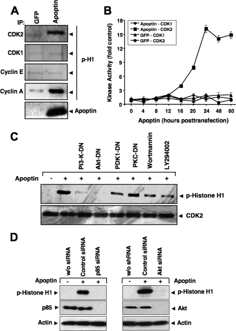
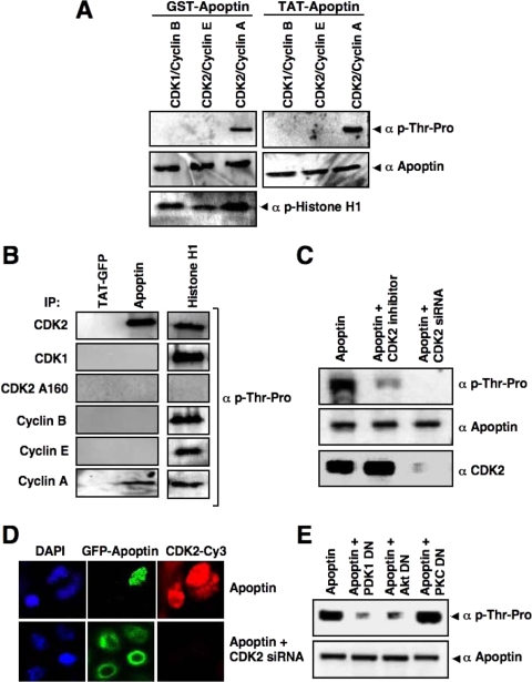
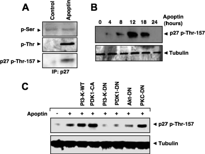
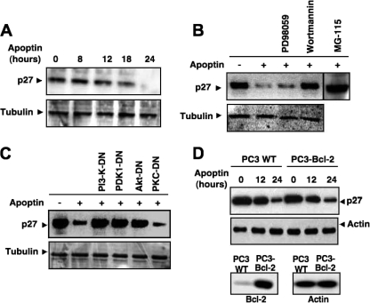
Apoptin, a protein from the chicken anemia virus, has attracted attention because it specifically kills tumor cells while leaving normal cells unharmed. The reason for this tumor selectivity is unclear and depends on subcellular localization, as apoptin resides in the cytoplasm of normal cells but in the nuclei of transformed cells. It was shown that nuclear localization and tumor-specific killing crucially require apoptin's phosphorylation by an as yet unknown kinase. Here we elucidate the pathway of apoptin-induced apoptosis and show that it essentially depends on abnormal phosphatidylinositol 3-kinase (PI3-kinase)/Akt activation, resulting in the activation of the cyclin-dependent kinase CDK2. Inhibitors as well as dominant-negative mutants of PI3-kinase and Akt not only inhibited CDK2 activation but also protected cells from apoptin-induced cell death. Akt activated CDK2 by direct phosphorylation as well as by the phosphorylation-induced degradation of the inhibitor p27(Kip1). Importantly, we also identified CDK2 as the principal kinase that phosphorylates apoptin and is crucially required for apoptin-induced cell death. Immortalized CDK2-deficient fibroblasts and CDK2 knockdown cells were markedly protected against apoptin. Thus, our results not only decipher the pathway of apoptin-induced cell death but also provide mechanistic insights for the selective killing of tumor cells.


Figures

FIG. 1.
FIG. 1.
Akt translocates to the nucleus during apoptin-induced cell death. (A) MCF-7 cells were transfected with GFP-tagged apoptin or a vector control. At 18 h posttransfection, the localization of Akt in either the absence or presence of apoptin in MCF-7 cells was detected by confocal microscopy after immunostaining with anti-Akt and Cy3-conjugated secondary antibody. DAPI (4′,6-diamidino-2-phenylindole) was used to counterstain nuclei, and the images were overlaid to determine the Akt localization within the cell. (B) MCF-7 cells were transfected with different apoptin mutants, and the localization of Akt and apoptin was detected by immunocytochemistry.
FIG. 2.
FIG. 2.
Apoptin interacts with Akt. (A) MCF-7 cells and HMECs were transfected with GFP-tagged apoptin. The interaction of endogenous Akt with apoptin was detected by immunoblotting with Akt antibody and immunoprecipitation with either control immunoglobulin G (IgG) or GFP antibody at 18 h posttransfection. (B) GST pull-down assay was performed using control GST or GST-apoptin immobilized on agarose beads and incubated with extracts prepared from either MCF-7 cells or HMECs. The interaction of Akt with apoptin was assessed by immunoblotting with anti-Akt antibody. (C and D) PC3 cells were infected with either adenoviral Myc-tagged NLS-Akt or a control virus in the presence or absence of GFP-apoptin. (C) Apoptotic cell death was measured by flow cytometric detection of hypodiploid DNA at 24 h or 48 h posttransfection. Data represent the means of four independent experiments. (D) The localization of NLS-Akt was detected after 18 h by anti-Myc-tag antibody followed by Cy3-marked secondary antibody. (E) Effect of NLS-Akt on apoptin-induced apoptosis in primary HMECs. Cells were transduced with an NLS-Akt or control virus as described for panel C. Cells were then treated with cell-permeative Tat-apoptin (1 μM) or left untreated. The percentage of apoptosis was determined after 30 h of treatment. (F) Apoptin triggers nuclear translocation of endogenous Akt in tumor cells but not in nontransformed cells. MCF-7 cells and HMECs were treated with Tat-apoptin for 18 h or left untreated. The differential localization of Akt was detected by immunostaining with anti-Akt and Cy3-conjugated secondary antibody. Nuclei were stained with DAPI.
FIG. 3.
FIG. 3.
CDK2 is activated during apoptin-induced cell death. (A) CDK2, CDK1, cyclin E, and cyclin A were immunoprecipitated from MCF-7 cells transfected with either GFP or GFP-apoptin at 24 h posttransfection and used in an in vitro kinase assay with histone H1 as the substrate. The level of histone H1 phosphorylation was detected by immunoblotting with a phospho-specific antibody against histone H1. (B) The kinase activities of CDK1 and CDK2 at different times after transfection with either GFP or apoptin were measured and plotted. Immunoblot signals were quantified against the respective controls, using a Storm scanner and accompanying software. (C) PC3 cells were transfected with apoptin alone or together with different dominant-negative mutants of PI3-K, Akt, PDK1, or PKCɛ. Kinase activity of immunoprecipitated CDK2 was measured using histone H1 as the substrate 24 h after transfection. Total CDK2 levels were determined by Western blotting. The PI3-K inhibitors wortmannin (5 nM) and LY294002 (1.5 μM) were applied 1 h before cell lysis. (D) Knockdown of PI3-K and Akt abolishes apoptin-induced CDK2 activation. PC3 cells were transfected with either control siRNA, a PI3-K p85-specific siRNA (left), or an Akt-specific siRNA (right). RNA interference resulted in an almost complete knockdown of p85 and Akt expression. At 72 h posttransfection, cells were treated with Tat-apoptin or left untreated. The kinase activity of immunoprecipitated CDK2 was determined by using histone H1 as the substrate 24 h after apoptin treatment. The level of histone H1 phosphorylation was detected by immunoblotting with a phospho-specific antibody against histone H1. Actin served as a loading control.
FIG. 4.
FIG. 4.
CDK2-cyclin A activity is required for apoptin-triggered cell death. (A) The effect of CDK2 inhibitor on apoptin's toxicity was measured by transfecting PC3 cells with GFP-apoptin in the presence or absence of the CDK2 inhibitor roscovitine (CDK2-I). Apoptosis was measured by flow cytometric detection of hypodiploid DNA after 24 and 48 h. (B) PC3 cells were transfected with plasmids for either CDK2 siRNA or control siRNA. After 48 h, CDK2 expression was detected by immunoblotting. (C) PC3 cells were transfected with GFP or GFP-apoptin and a CDK2-specific or control siRNA plasmid. Apoptosis was quantified after 24 and 48 h by measurement of the formation of hypodiploid DNA. (D) Immortalized CDK2-deficient murine fibroblasts and the respective WT control cells were treated with cell-permeative Tat-apoptin (1 μM) for either 24 or 48 h, and apoptosis was then evaluated by flow cytometry. Data represent the averages of four independent experiments. In both cell populations, cell death in the absence of apoptin did not exceed 4%. (E) CDK2 kinase activity was measured 24 h after transfection of PC3 cells with apoptin alone, with cotreatment with the CDK2 inhibitor roscovitine (CDK2-I) or the caspase inhibitor Z-VAD-FMK, or with overexpression of Bcl-2. (F) Levels of cytosolic cytochrome c were detected by cellular fractionation and immunoblotting with PC3 cells transfected with apoptin alone, with cotreatment with CDK2 inhibitor, or with Bcl-2 overexpression. Actin served as a loading control.
FIG. 5.
FIG. 5.
CDK2 is the specific apoptin kinase. (A) A nonradioactive in vitro kinase assay was performed with recombinant GST-apoptin and Tat-apoptin as substrates, using active CDK1-cyclin B, CDK2-cyclin E, or CDK2-cyclin A. Apoptin phosphorylation was detected by immunoblotting using an antibody against phosphorylated threonine-proline residues. Total apoptin levels were detected by apoptin antibody. Histone H1 was used as a positive control. (B) Active CDK2, CDK1, cyclin B, cyclin E, and cyclin A were immunoprecipitated from PC3 cells with their respective antibodies and the CDK2 T160A mutant was immunoprecipitated with anti-hemagglutinin antibody, and the immunoprecipitates were used in a kinase assay with Tat-GFP, Tat-apoptin, or histone H1 as the substrate. Phosphorylation was detected as described in the legend to Fig. 4A. (C) PC3 cells were transfected with GFP-apoptin alone or in the presence of the CDK2 inhibitor or the CDK2-targeting siRNA. GFP-apoptin was immunoprecipitated at 24 h posttransfection with anti-GFP antibodies. The phosphorylation of apoptin in the immunoprecipitates was detected by immunoblotting using anti-phospho-Thr-Pro antibodies. Total apoptin and CDK2 levels are indicated. (D) The effect of CDK2 inhibition on apoptin's localization was demonstrated in PC3 cells transfected with GFP-apoptin in the absence or presence of a CDK2-targeting siRNA plasmid. After 24 h, cells were analyzed by confocal laser scanning microscopy. (E) PI3-K/Akt signaling is required for apoptin phosphorylation. PC3 cells were transfected with GFP-apoptin in the presence or absence of a dominant-negative mutant of PDK1, Akt, or PKCɛ. The levels of apoptin phosphorylation were determined at 24 h posttransfection as described for panel C. Total apoptin levels were detected with anti-apoptin antibodies.
FIG. 6.
FIG. 6.
p27Kip1 is phosphorylated during apoptin-induced cell death. (A) PC3 cells were transfected with the GFP control or GFP-apoptin. After 12 h, the phosphorylation of p27Kip1 was detected by phospho-Ser- or phospho-Thr-specific antibodies after immunoprecipitation with anti-p27Kip1 antibody. Threonine phosphorylation was also detected with an antibody against Thr-157-phosphorylated p27Kip1. (B) p27Kip1 phosphorylation after apoptin transfection at different time points was detected by immunoblotting with phospho-Thr-157-specific p27Kip1 antibody. Tubulin was used as a loading control. (C) Cells were transfected with apoptin alone or cotransfected with either WT PI3-K, constitutively active PDK1 (CA), PI3-K dominant-negative (DN) vector, PDK1-DN, Akt-DN, or PKCɛ-DN vector. After 16 h, cells were lysed and phosphorylated p27Kip1 was detected by Western blotting.
FIG. 7.
FIG. 7.
Akt-mediated phosphorylation enhances degradation of p27Kip1 via the proteasomal pathway. (A) Levels of p27Kip1 in GFP-apoptin-transfected PC3 cells were monitored for 24 h posttransfection by Western blotting. Tubulin was used as a loading control. (B) PC3 cells, transfected with apoptin or left untransfected, were incubated with the MEK inhibitor PD98059, the PI3-K inhibitor wortmannin, or the proteasomal inhibitor MG115. Protein levels of p27Kip1 were detected by immunoblotting. (C) Roles of PI3-K and Akt in p27Kip1 stability. Protein levels of p27Kip1 were detected by immunoblotting of lysates of PC3 cells 24 h after transfection with apoptin and the indicated dominant-negative mutant of PI3-K, Akt, PDK1, or PKCɛ. (D) Bcl-2 does not affect degradation of p27Kip1. WT and Bcl-2-overexpressing PC3 cells were treated with Tat-apoptin for the indicated times, and the protein level of p27Kip1 was evaluated by immunoblotting. Actin was used as a loading control. The status of Bcl-2 expression and expression of an actin control in the two cell lines are shown in the lower panels.
FIG. 8.
FIG. 8.
p27Kip1 is involved in apoptin-induced apoptosis. (A) Downregulation of p27Kip1 protects against apoptin-induced apoptosis. PC3 cells were transfected with a GFP control or GFP-apoptin in the presence and absence of a p27Kip1-specific siRNA. (Left) Apoptosis was measured by flow cytometric detection of hypodiploid DNA at the indicated time points. The data represent the means from three independent experiments. (Right) The efficiency of p27Kip1 downregulation by the specific or scrambled control siRNA was investigated at 36 h posttransfection by immunoblotting with a p27Kip1-specific antibody. Tubulin served as a loading control. (B to D) Apoptin-induced apoptosis is inhibited in cells with suppressed expression of Skp2 and Cks1, two components of the p27 degradation machinery. (B) PC3 cells were transfected with either a control siRNA, two different Skp2 siRNAs (left panels), or two different Cks1 siRNAs (right panels). The protein levels of Skp2, Cks1, p27, and actin were analyzed by immunoblotting at 72 h post-siRNA transfection. (C) PC3 cells transfected with either control siRNA, Skp2 siRNA no. 2, or Cks1 siRNA no. 2 were treated with Tat-apoptin (1 μM) at 36 h posttransfection, and the percentage of apoptosis was measured after 24 h of further incubation. (D) Immortalized Skp2-deficient murine fibroblasts and the respective WT control cells were treated with Tat-apoptin, and the percentage of apoptosis was evaluated after 24 and 48 h of treatment. Data represent the averages of three independent experiments.
FIG. 9.
FIG. 9.
Model for apoptin-activated signaling. Apoptin interacts with the SH3 domain of PI3-K, resulting in its constitutive activation, which leads to PDK1-dependent Akt activation and nuclear translocation of Akt. Nuclear Akt activates CDK2 by both direct and indirect mechanisms, including the proteasome-dependent degradation of p27Kip1. The activated CDK2 phosphorylates, among other substrates, apoptin at Thr-108 and thereby enforces its nuclear accumulation in cancer cells. The abnormal CDK2 activation, which might also involve apoptin-induced inhibition of APC/C, directly or indirectly influences the activity of regulators of the mitochondrial apoptotic pathway, including Nur77, Bcl-2, Bim, and others.

Similar articles

Cited by

References

    1. Ahn, J. Y., X. Liu, Z. Liu, L. Pereira, D. Cheng, J. Peng, P. A. Wade, A. W. Hamburger, and K. Ye. 2006. Nuclear Akt associates with PKC-phosphorylated Ebp1, preventing DNA fragmentation by inhibition of caspase-activated DNase. EMBO J. 252083-2095. - PMC - PubMed
    1. Aki, T., K. Yamaguchi, T. Fujimiya, and Y. Mizukami. 2003. Phosphoinositide 3-kinase accelerates autophagic cell death during glucose deprivation in the rat cardiomyocyte-derived cell line H9c2. Oncogene 228529-8535. - PubMed
    1. Backendorf, C., A. E. Visser, A. G. de Boer, R. Zimmerman, M. Visser, P. Voskamp, Y. H. Zhang, and M. H. Noteborn. 2008. Apoptin: therapeutic potential of an early sensor of carcinogenic transformation. Annu. Rev. Pharmacol. Toxicol. 48143-169. - PubMed
    1. Barkett, M., and T. D. Gilmore 1999. Control of apoptosis by Rel/NF-kappaB transcription factors. Oncogene 186910-6924. - PubMed
    1. Bashir, T., N. V. Dorrello, V. Amador, D. Guardavaccaro, and M. Pagano. 2004. Control of the SCF(Skp2-Cks1) ubiquitin ligase by the APC/C(Cdh1) ubiquitin ligase. Nature 428190-193. - PubMed

Publication types

Substances

LinkOut - more resources