. 2009 Mar;77(3):1230-7.
doi: 10.1128/IAI.01117-08. Epub 2008 Dec 22.

Role of the dosR-dosS two-component regulatory system in Mycobacterium tuberculosis virulence in three animal models

Affiliations

Role of the dosR-dosS two-component regulatory system in Mycobacterium tuberculosis virulence in three animal models

Paul J Converse et al. Infect Immun. 2009 Mar.

Abstract

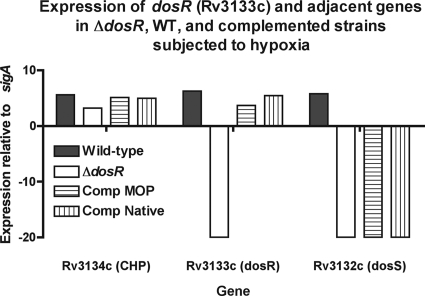
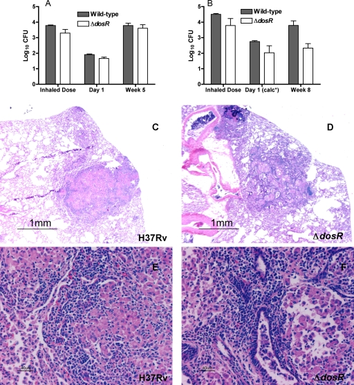
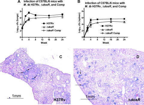
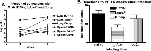
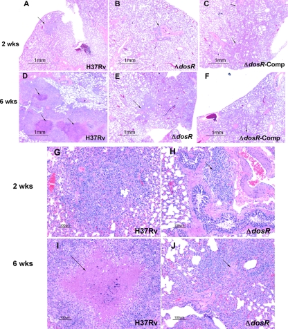
The Mycobacterium tuberculosis dosR gene (Rv3133c) is part of an operon, Rv3134c-Rv3132c, and encodes a response regulator that has been shown to be upregulated by hypoxia and other in vitro stress conditions and may be important for bacterial survival within granulomatous lesions found in tuberculosis. DosR is activated in response to hypoxia and nitric oxide by DosS (Rv3132c) or DosT (Rv2027c). We compared the virulence levels of an M. tuberculosis dosR-dosS deletion mutant (DeltadosR-dosS [DeltadosR-S]), a dosR-complemented strain, and wild-type H37Rv in rabbits, guinea pigs, and mice infected by the aerosol route and in a mouse hollow-fiber model that may mimic in vivo granulomatous conditions. In the mouse and the guinea pig models, the DeltadosR-S mutant exhibited a growth defect. In the rabbit, the DeltadosR-S mutant did not replicate more than the wild type. In the hollow-fiber model, the mutant phenotype was not different from that of the wild-type strain. Our analyses reveal that the dosR and dosS genes are required for full virulence and that there may be differences in the patterns of attenuation of this mutant between the animal models studied.


Figures

FIG. 1.
FIG. 1.
Expression of components of the dosR operon (Rv3134c-Rv3132c). Four strains, the H37Rv wild-type (WT) parental strain (solid bars), a putative dosR deletion mutant, and two ΔdosR-complemented strains with native and mycobacterial optimal promoters (MOP), were subjected to hypoxic stress (see Materials and Methods) to enhance expression. Extracted DNA-free RNA was subjected to RT, and the cDNA was subjected to 40 cycles of quantitative PCR. Expression was normalized to that of the housekeeping gene sigA (∼20 cycles), such that enhanced expression (fewer cycles to detection [CT]) is shown as being positive and those with undetectable product are shown as being negative. The deletion mutant has no detectable dosR or dosS, while the complemented strains have restored dosR but not dosS.
FIG. 2.
FIG. 2.
Comparison of M. tuberculosis H37Rv:ΔdosR-S and wild-type H37Rv in rabbits after aerosol infection. In a first 5-week experiment (A), the bacterial burden was comparable at 5 weeks, while in a second 8-week experiment (B), there was a significantly (P = 0.0312 by Wilcoxon test) lower bacillary burden at 8 weeks. The average inhaled doses in each experiment were not significantly different between bacterial strains. The differences were also not significant with regard to the number of bacteria implanted (1.91 ± 0.04 log10 CFU for the wild type and 1.66 ± 0.097 log10 CFU for the ΔdosR-S strain) in the 5-week experiment (A) and were also not significant in the 8-week experiment (B), in which the doses implanted at day 1 were calculated (calc*) based on the differences between inhaled and implanted doses in the first experiment, which are similar to those reported previously in the literature for the rabbit model (8, 18). For a detailed explanation, see the text. Histopathological analysis of rabbit lungs 5 weeks after infection with M. tuberculosis H37Rv (C and E) shows a well-organized primary granulomatous lesion with dark lymphocytes surrounding pale epithelioid macrophages capable of killing tubercle bacilli. In contrast, with the ΔdosR-S deletion mutant (D and F), the lesions were fewer and smaller. Magnifications, ×20 (C and D) and ×200 (E and F).
FIG. 3.
FIG. 3.
Growth after well-matched implantation of wild-type H37Rv, ΔdosR-S, and Comp strains. In spite of comparable implantation levels, the ΔdosR-S mutant did not multiply as well in the lungs (A) or the spleen (B) as the wild type. The ΔdosR-S mutant complemented with the mycobacterial optimal promoter (Comp) does not grow as well in the lungs or spleen as the wild-type strain even with comparable levels of implantation (2.71 ± 0.19 log10 CFU for H37Rv, 3.11 ± 0.11 log10 CFU for the ΔdosR-S strain, and 2.98 ± 0.20 log10 CFU for Comp) over the first 28 days. (C and D) Histopathology of lungs of C57BL/6 mice infected with M. tuberculosis (M. tb) H37Rv (C) or a ΔdosR-S deletion mutant (D) 2 months after infection. At 2 months, the lesions were large and dense in wild-type-infected mouse lungs compared to the scattered lesions in ΔdosR-S-infected mouse lungs, in which more normal lung tissue was preserved. No lesions showed evidence of necrosis. Magnification, ×20.
FIG. 4.
FIG. 4.
Guinea pigs resist multiplication of M. tuberculosis (M. tb) cells lacking dosR and dosS. (A) After infection with approximately 1.3 log10 CFU, the complemented strain (Comp) initially multiplied at an intermediate level between H37Rv wild-type and ΔdosR-S mutant bacillus levels before the onset of tuberculin reactivity, but by week 6, the growth of this strain declined to the same level as that seen for the ΔdosR-S mutant bacilli. Lungs are shown with solid lines, and spleens are shown with dashed lines. (B) Reactivity to tuberculin (PPD). The wild-type and complemented (Comp) strains elicit a stronger (P < 0.025 by one-way analysis of variance) mean delayed-type hypersensitivity response to PPD than does the ΔdosR-S mutant (horizontal stripes).
FIG. 5.
FIG. 5.
(A to C) Histopathology of lungs of guinea pigs infected with M. tuberculosis H37Rv, a ΔdosR-S deletion mutant, or the mutant complemented with dosR at 2 weeks, before the onset of tuberculin reactivity. Only a small early granulomatous, noncaseating lesion (arrow) in the lung of a wild-type-infected guinea pig was observed at this time (A). Mutant- and complement-infected guinea pigs showed only rare inflammatory peribronchial/periarteriolar infiltrates (arrows). (D to F) At 6 weeks, there was strong tuberculin reactivity (see Table S2 in the supplemental material) and substantial inflammation and necrosis (arrows) in the lungs of wild-type-infected guinea pigs (D), almost none detectable in the mutant-infected animals (E), and small, poorly organized granulomas (arrows) in the complement-infected guinea pigs (F). Magnification, ×20. (G to J) Above-described lung lesions of guinea pigs infected with H37Rv wild-type (G and I) and ΔdosR-S (H and J) mutant strains at higher magnifications. Caseous necrosis (arrow) is apparent in the lesions of the wild-type-infected guinea pig at 6 weeks (I), whereas a rare lesion (J) in a ΔdosR-S mutant-infected animal shows rather poor organization and limited necrosis (arrow). Magnification, ×100.

References

    1. Abdul-Majid, K.-B., L. Ly, P. Converse, D. Geiman, D. McMurray, and W. Bishai. 2008. Altered cellular infiltration and cytokine levels during early Mycobacterium tuberculosis sigC mutant infection are associated with late-stage disease attenuation and milder immunopathology in mice. BMC Microbiol. 8151. - PMC - PubMed
    1. Aly, S., K. Wagner, C. Keller, S. Malm, A. Malzan, S. Brandau, F. C. Bange, and S. Ehlers. 2006. Oxygen status of lung granulomas in Mycobacterium tuberculosis-infected mice. J. Pathol. 210298-305. - PubMed
    1. Boon, C., and T. Dick. 2002. Mycobacterium bovis BCG response regulator essential for hypoxic dormancy. J. Bacteriol. 1846760-6767. - PMC - PubMed
    1. Boshoff, H. I. M., and C. E. Barry. 2005. Tuberculosis metabolism and respiration in the absence of growth. Nat. Rev. Microbiol. 370-80. - PubMed
    1. Chan, J., K. Tanaka, D. Carroll, J. Flynn, and B. R. Bloom. 1995. Effects of nitric oxide synthase inhibitors on murine infection with Mycobacterium tuberculosis. Infect. Immun. 63736-740. - PMC - PubMed

Publication types

MeSH terms

Substances