. 2008 Dec 29;183(7):1235-42.
doi: 10.1083/jcb.200809113. Epub 2008 Dec 22.

The sympathetic tone mediates leptin's inhibition of insulin secretion by modulating osteocalcin bioactivity

Affiliations

The sympathetic tone mediates leptin's inhibition of insulin secretion by modulating osteocalcin bioactivity

Eiichi Hinoi et al. J Cell Biol. .

Abstract

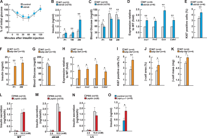
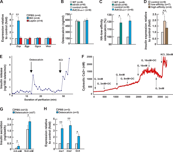
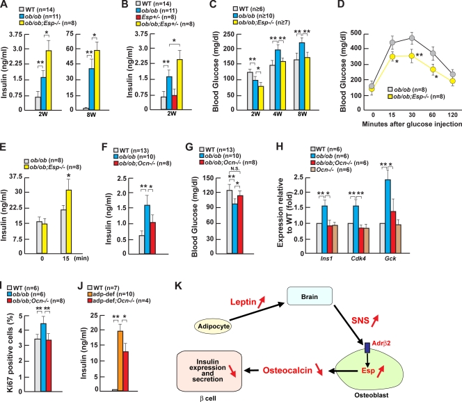
The osteoblast-secreted molecule osteocalcin favors insulin secretion, but how this function is regulated in vivo by extracellular signals is for now unknown. In this study, we show that leptin, which instead inhibits insulin secretion, partly uses the sympathetic nervous system to fulfill this function. Remarkably, for our purpose, an osteoblast-specific ablation of sympathetic signaling results in a leptin-dependent hyperinsulinemia. In osteoblasts, sympathetic tone stimulates expression of Esp, a gene inhibiting the activity of osteocalcin, which is an insulin secretagogue. Accordingly, Esp inactivation doubles hyperinsulinemia and delays glucose intolerance in ob/ob mice, whereas Osteocalcin inactivation halves their hyperinsulinemia. By showing that leptin inhibits insulin secretion by decreasing osteocalcin bioactivity, this study illustrates the importance of the relationship existing between fat and skeleton for the regulation of glucose homeostasis.


Figures

Figure 1.
Figure 1.
Leptin regulates insulin secretion in part through the neuronal pathway. (A) ITT in 2-wk-old ob/ob mice. (B and C) Serum insulin and blood glucose in ob/ob mice. (D) Gene expression in pancreas or islets of 2-wk-old ob/ob mice. (E) Quantification of insulin/Ki67 immunoreactive cells in islets of 2-wk-old ob/ob mice. (F and G) Serum insulin and blood glucose in adipocyte-deficient (adp-def) mice. (H) Gene expression in pancreas or islets of 2-wk-old adipocyte-deficient mice. (I–K) Quantification of insulin/Ki67 immunoreactive cells in islets, β-cell area, and β-cell mass of 2-wk-old adipocyte-deficient mice. (L–N) Glucose-stimulated insulin secretion by leptin in islets from WT, ob/ob, and L/L mice. (O) Serum insulin levels in 1-mo-old Leprsyn−/− mice. Error bars indicate mean + SEM. *, P < 0.05; **, P < 0.01; P1, newborn; 1W, 1 wk old; 2W, 2 wk old. Control in O indicates Synapsin-Cre mice. In L–N, the concentration of glucose in the culture media is indicated in millimolars.
Figure 2.
Figure 2.
Sympathetic signaling in osteoblasts regulates insulin expression and secretion. (A) Ucp1 expression in brown fat of Leprsyn−/− mice. (B and C) Serum epinephrine and norepinephrine levels in Leprsyn−/− mice. (D and E) Serum insulin levels in ob/ob and adipocyte-deficient (adp-def) mice after daily isoproterenol (+ISO), phenylephrine (+Phe), or clonidine (+Clo) injection for 2 wk. (F) Ucp1 expression in brown fat of ob/ob and adipocyte-deficient mice after daily isoproterenol injection. (G) Serum insulin levels in 2-wk-old Adrβ2osb−/− mice. (H) Serum insulin levels in WT and Adrβ2osb−/− mice after daily propranolol injection for 2 wk. (I and J) Blood glucose levels and gene expression in islets of 2-wk-old Adrβ2osb−/− mice. (K–M) Serum insulin, blood glucose, and Insulin expression in the pancreas of 2-wk-old ob/+;Adrβ2osb+/− mice. (N) Delta insulin after glucose-stimulated insulin secretion in WT and Adrβ2osb−/− mice treated with central leptin infusion at 4 ng/h for 1 wk. (O and P) Plasma insulin levels during hyperglycemic clamps in WT and Adrβ2osb−/− mice treated with central leptin infusion at 4 ng/h for 1 wk. Error bars indicate mean + SEM. *, P < 0.05; **, P < 0.01. Control in A–C indicates Synapsin-Cre mice. Control in G–J and M indicates α1(I)Collagen-Cre mice.
Figure 3.
Figure 3.
Osteocalcin directly regulates insulin secretion in a Ca2+-dependent manner. (A) Gene expression in isoproterenol (ISO)- or leptin-treated osteoblasts. (B and C) Levels of total and uncarboxylated osteocalcin (HA low affinity) in ob/ob and Adrβ2osb−/− mice. (D) Insulin expression by HA affinity fraction in islets. (E) Perifusion of WT islets in the presence of 0.03 ng/ml osteocalcin under a 3-mM glucose condition. (F) Intracellular calcium imaging assay using WT islets in the presence of osteocalcin under the different concentrations of glucose. (G) Insulin secretion by osteocalcin in WT islets maintained in either low or high glucose. (H) Gene expression in osteocalcin-treated islets. Error bars indicate mean + SEM. *, P < 0.05; **, P < 0.01. Control in B and C indicates α1(I)Collagen-Cre mice. In G, the concentration of glucose in the culture media is indicated in millimolars.
Figure 4.
Figure 4.
Leptin regulates insulin secretion through osteoblasts. (A) Serum insulin in 2- and 8-wk-old ob/ob;Esp−/− mice. (B) Serum insulin in 2-wk-old ob/ob;Esp−/− mice. (C) Blood glucose in 2–8-wk-old ob/ob;Esp−/− mice. (D) Glucose tolerance tests in 2-mo-old ob/ob;Esp−/− mice. (E) Serum insulin after glucose challenge in 2-mo-old ob/ob;Esp−/− mice. (F and G) Serum insulin and blood glucose in 2-wk-old ob/ob;Ocn−/− mice. (H) Gene expression in pancreas or islets of 2-wk-old ob/ob;Ocn−/− mice. (I) Quantification of insulin/Ki67-positive cell islets of 2-wk-old ob/ob;Ocn−/− mice. (J) Serum insulin in 2-wk-old adipocyte-deficient;Ocn−/− mice. adp-def, adipocyte deficient. (K) In the presence of leptin, sympathetic tone is high; thus, Esp expression in the osteoblast is high, and osteocalcin bioactivity is low. This contributes to the inhibition of insulin secretion by leptin. Error bars indicate mean + SEM. *, P < 0.05; **, P < 0.01. 2W, 2 wk old; 4W, 4 wk old; 8W, 8 wk old; SNS, sympathetic nervous system.

References

    1. Altman, J.D., A.U. Trendelenburg, L. MacMillan, D. Bernstein, L. Limbird, K. Starke, B.K. Kobilka, and L. Hein. 1999. Abnormal regulation of the sympathetic nervous system in alpha2A-adrenergic receptor knockout mice. Mol. Pharmacol. 56:154–161. - PubMed
    1. Berthoud, H.R., and B. Jeanrenaud. 1979. Acute hyperinsulinemia and its reversal by vagotomy after lesions of the ventromedial hypothalamus in anesthetized rats. Endocrinology. 105:146–151. - PubMed
    1. Björnholm, M., H. Münzberg, R.L. Leshan, E.C. Villanueva, S.H. Bates, G.W. Louis, J.C. Jones, R. Ishida-Takahashi, C. Bjørbaek, and M.G. Myers Jr. 2007. Mice lacking inhibitory leptin receptor signals are lean with normal endocrine function. J. Clin. Invest. 117:1354–1360. - PMC - PubMed
    1. Bügel, S. 2008. Vitamin K and bone health in adult humans. Vitam. Horm. 78:393–416. - PubMed
    1. Cho, Y.R., H.J. Kim, S.Y. Park, H.J. Ko, E.G. Hong, T. Higashimori, Z. Zhang, D.Y. Jung, M.S. Ola, K.F. Lanoue, et al. 2007. Hyperglycemia, maturity-onset obesity, and insulin resistance in NONcNZO10/LtJ males, a new mouse model of type 2 diabetes. Am. J. Physiol. Endocrinol. Metab. 293:E327–E336. - PubMed

Publication types