. 2009 Jan 16;206(1):113-23.
doi: 10.1084/jem.20081244. Epub 2008 Dec 22.

Absence of SPARC results in increased cardiac rupture and dysfunction after acute myocardial infarction

Affiliations

Absence of SPARC results in increased cardiac rupture and dysfunction after acute myocardial infarction

Mark W M Schellings et al. J Exp Med. .

Abstract

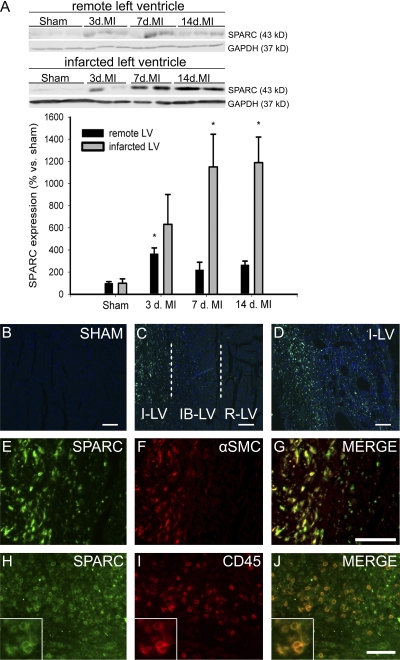
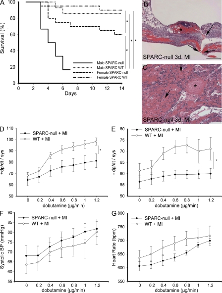
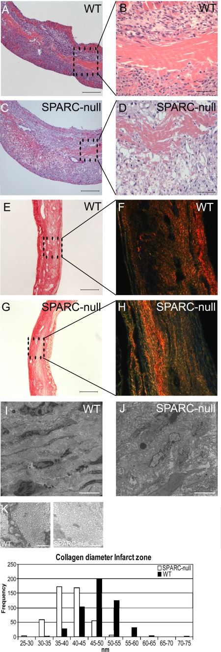
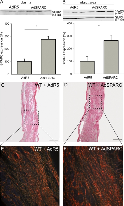
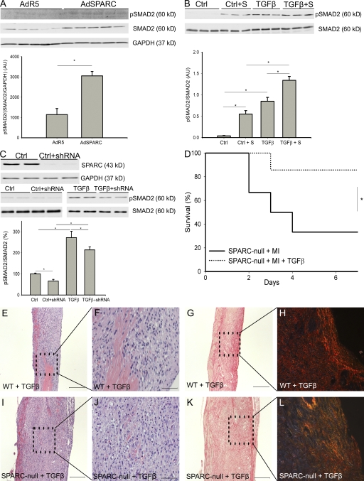
The matricellular protein SPARC (secreted protein, acidic and rich in cysteine, also known as osteonectin) mediates cell-matrix interactions during wound healing and regulates the production and/or assembly of the extracellular matrix (ECM). This study investigated whether SPARC functions in infarct healing and ECM maturation after myocardial infarction (MI). In comparison with wild-type (WT) mice, animals with a targeted inactivation of SPARC exhibited a fourfold increase in mortality that resulted from an increased incidence of cardiac rupture and failure after MI. SPARC-null infarcts had a disorganized granulation tissue and immature collagenous ECM. In contrast, adenoviral overexpression of SPARC in WT mice improved the collagen maturation and prevented cardiac dilatation and dysfunction after MI. In cardiac fibroblasts in vitro, reduction of SPARC by short hairpin RNA attenuated transforming growth factor beta (TGF)-mediated increase of Smad2 phosphorylation, whereas addition of recombinant SPARC increased Smad2 phosphorylation concordant with increased Smad2 phosphorylation in SPARC-treated mice. Importantly, infusion of TGF-beta rescued cardiac rupture in SPARC-null mice but did not significantly alter infarct healing in WT mice. These findings indicate that local production of SPARC is essential for maintenance of the integrity of cardiac ECM after MI. The protective effects of SPARC emphasize the potential therapeutic applications of this protein to prevent cardiac dilatation and dysfunction after MI.


Figures

Figure 1.
Figure 1.
SPARC expression is induced after MI. (A) SPARC protein is increased after MI in mice. Representative Western blots of SPARC in remote and infarcted LV from WT mice at 3, 7, and 14 d after MI (n = 4 per time point; *, P < 0.05). (B–D) SPARC immunofluorescent staining (green) is absent in sham-operated hearts (B) and remote LV (R-LV), gradually increases in the infarct border LV (IB-LV; C), and is strongly up-regulated in the infarcted LV (I-LV) 7 d after MI (C and D). (E–J) SPARC expression (E and H) colocalizes with α–smooth muscle cell (SMC) actin–positive myofibroblasts (F and G) and CD45 immunoreactive leukocytes (I and J) in the infarcted LV of WT infarcted hearts 7 d after MI. The insets in H–J show detailed SPARC and CD45 immunoreactive leukocytes. Error bars represent the mean ± SEM. Bars: (B–D, H, and J) 100 μm; (E–G) 50 μm.
Figure 2.
Figure 2.
Absence of SPARC results in cardiac rupture and dysfunction. (A) Kaplan-Meier curve showing that targeted deletion of SPARC resulted in decreased survival of SPARC-null (mainly male) compared with WT mice after MI (*, P < 0.05). Decreased survival was mainly caused by cardiac rupture. Two male SPARC-null mice had to be killed at 7 d after MI because of severe shortness of breath and, therefore, were not included in the survival curve. (B and C) Histological analysis of ruptured LV of male SPARC-null infarcted hearts (hematoxylin and eosin stained) revealing rupture site (B, arrows), intramural hemorrhages of the infarcted ventricular wall (C, arrows), and massive infiltration of erythrocytes and inflammatory cells (C) and thrombi (B and C, asterisks) at 3 d after MI. (D–G) Decreased survival is associated with depressed cardiac contractility (D) and relaxation (E) during infusion of dobutamine in SPARC-null female (n = 7) in comparison with female WT (n = 9) mice 14 d after MI, whereas systolic blood pressure (BP; F) and heart rate (G) did not differ significantly (*, P < 0.05). bpm, beats per minute. Error bars represent the mean ± SEM. Bars: (B) 200 μm; (C) 100 μm.
Figure 3.
Figure 3.
Adverse wound healing in SPARC-null mice. (A–D) Hematoxylin and eosin staining revealed a disorganized granulation tissue with increased RBC infiltration in male SPARC-null (C and D) versus WT (A and B) 7-d-old infarcts. (E–K) Abnormal collagen formation in SPARC-null infarcts. (E–H) Sirius red staining did not indicate substantial differences in the amount of collagen deposition between WT (E) and SPARC-null (G) infarcts at 14 d after MI. Sirius red polarization microscopy revealed well aligned and tightly packed (orange-red) collagen fibers in WT (F) but less mature fibers (yellow-green) in SPARC-null infarcts (H). (I–K) Ultrastructural analysis confirmed a disorganized matrix in SPARC-null (J) in comparison with WT (I) infarcts, which was associated with the deposition of smaller collagen fibrils in the infarct zone of SPARC-null mice (K; n = 3). Bars: (A, C, E, and G) 200 μm; (B and D) 50 μm; (I and J) 5 μm; (K) 500 nm.
Figure 4.
Figure 4.
SPARC overexpression increases collagen deposition and quality after MI. (A and B) Representative Western blots showing a 2.8-fold increase of SPARC in plasma (A) and a 2.6-fold increase of SPARC in the infarct area (B) of AdSPARC-treated WT mice in comparison with AdR5-treated WT mice at 14 d after MI (n = 4; *, P < 0.05). (C–F) Sirius red staining and polarization microscopy revealed increased collagen deposition in AdSPARC-treated (D) in comparison with AdR5-treated WT infarcts (C) and more well aligned and tightly packed (orange-red) collagen fibers in AdSPARC-treated (F) in comparison with AdR5-treated WT infarcts (E). Error bars represent the mean ± SEM. Bars: (C and D) 2 mm; (E and F) 200 μm.
Figure 5.
Figure 5.
SPARC and TGF-β cooperate during infarct healing. (A) Representative Western blot showing increased phosphorylated and total Smad2 levels in the infarcts of AdSPARC-treated mice (n = 9; *, P < 0.05). (B) Representative Western blot showing that addition of recombinant mouse SPARC activated Smad2 signaling and augmented the Smad2-activating potential of TGF-β (n = 6; *, P < 0.05). (C) The top blot is a representative Western blot indicating that shRNA against SPARC resulted in an 80% decrease in levels of SPARC protein. The blots are representative Western blots showing that Smad2 phosphorylation in shRNA-treated cardiac fibroblasts was significantly decreased at baseline levels (left) and after 15 min of stimulation by 1 ng/ml TGF-β (right; n = 5 per group; *, P < 0.05). (D) Survival curve showing that infusion of TGF-β protected against cardiac rupture after MI in male SPARC-null mice (1 out of 7 mice) in comparison with saline-treated male SPARC-null mice (8 out of 12 mice; *, P < 0.05). (E, F, I, and J) Hematoxylin and eosin staining revealed striking differences between TGF-β–treated WT and SPARC-null mice 7 d after MI. In WT mice, infusion of TGF-β did not significantly affect wound healing, whereas in SPARC-null animals it stimulated ECM production in the infarct zone. (G, H, K, and L) Sirius Red staining and Sirius Red polarization microscopy revealed no apparent differences in collagen deposition between TGF-β1–treated SPARC-null and WT mice, whereas the difference in well aligned and tightly packed (orange-red) collagen fibers in WT infarcts (H), but less mature fibers (yellow-green) in SPARC-null infarcts (L), was preserved. Error bars represent the mean ± SEM. Bars: (E, G–I, K, and L) 200 μm; (F and J) 50 μm.

References

    1. Framson, P.E., and E.H. Sage. 2004. SPARC and tumor growth: where the seed meets the soil? J. Cell. Biochem. 92:679–690. - PubMed
    1. Bradshaw, A.D., and E.H. Sage. 2001. SPARC, a matricellular protein that functions in cellular differentiation and tissue response to injury. J. Clin. Invest. 107:1049–1054. - PMC - PubMed
    1. Dobaczewski, M., M. Bujak, P. Zymek, G. Ren, M.L. Entman, and N.G. Frangogiannis. 2006. Extracellular matrix remodeling in canine and mouse myocardial infarcts. Cell Tissue Res. 324:475–488. - PubMed
    1. Komatsubara, I., T. Murakami, S. Kusachi, K. Nakamura, S. Hirohata, J. Hayashi, S. Takemoto, C. Suezawa, Y. Ninomiya, and Y. Shiratori. 2003. Spatially and temporally different expression of osteonectin and osteopontin in the infarct zone of experimentally induced myocardial infarction in rats. Cardiovasc. Pathol. 12:186–194. - PubMed
    1. Basu, A., L.H. Kligman, S.J. Samulewicz, and C.C. Howe. 2001. Impaired wound healing in mice deficient in a matricellular protein SPARC (osteonectin, BM-40). BMC Cell Biol. 2:15. - PMC - PubMed

Publication types

MeSH terms