. 2009 Jan 6;119(1):99-106.
doi: 10.1161/CIRCULATIONAHA.108.799700. Epub 2008 Dec 22.

Acute doxorubicin cardiotoxicity is associated with p53-induced inhibition of the mammalian target of rapamycin pathway

Affiliations

Acute doxorubicin cardiotoxicity is associated with p53-induced inhibition of the mammalian target of rapamycin pathway

Wuqiang Zhu et al. Circulation. .

Abstract

Background: Doxorubicin is used to treat childhood and adult cancer. Doxorubicin treatment is associated with both acute and chronic cardiotoxicity. The cardiotoxic effects of doxorubicin are cumulative, which limits its chemotherapeutic dose. Free radical generation and p53-dependent apoptosis are thought to contribute to doxorubicin-induced cardiotoxicity.

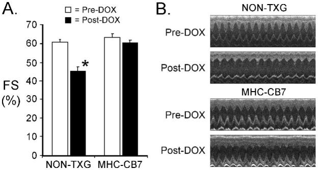
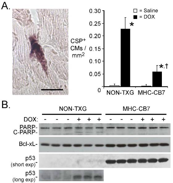
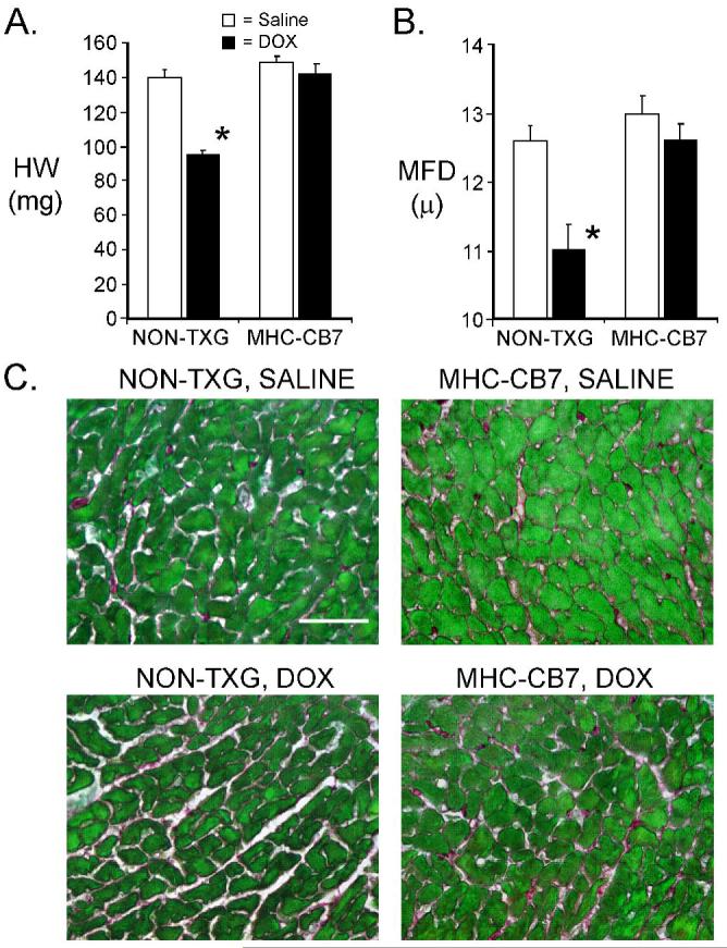
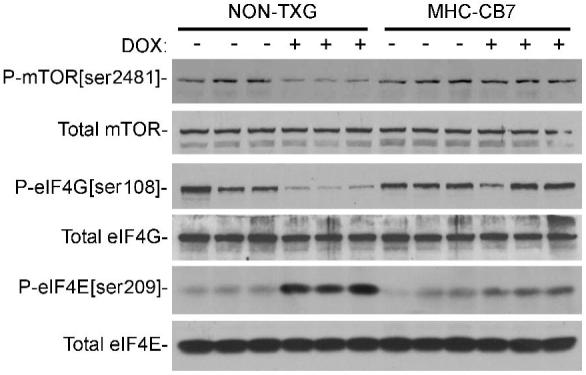
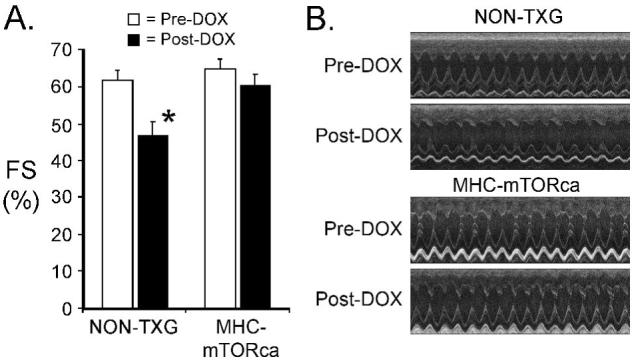
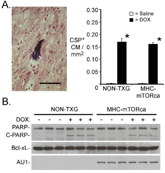
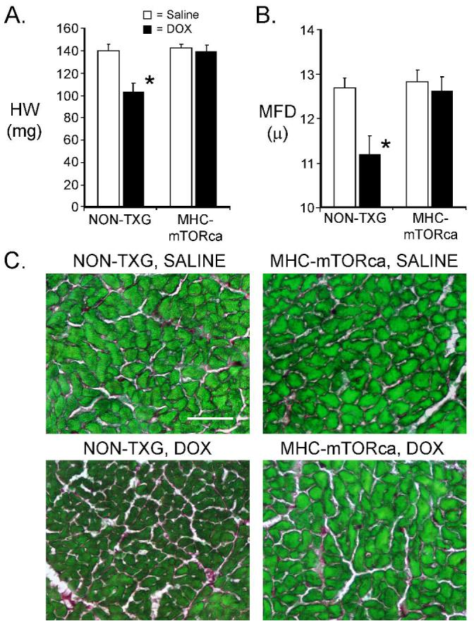
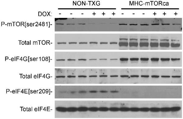
Methods and results: Adult transgenic (MHC-CB7) mice expressing cardiomyocyte-restricted dominant-interfering p53 and their nontransgenic littermates were treated with doxorubicin (20 mg/kg cumulative dose). Nontransgenic mice exhibited reduced left ventricular systolic function (predoxorubicin fractional shortening [FS] 61+/-2%, postdoxorubicin FS 45+/-2%, mean+/-SEM, P<0.008), reduced cardiac mass, and high levels of cardiomyocyte apoptosis 7 days after the initiation of doxorubicin treatment. In contrast, doxorubicin-treated MHC-CB7 mice exhibited normal left ventricular systolic function (predoxorubicin FS 63+/-2%, postdoxorubicin FS 60+/-2%, P>0.008), normal cardiac mass, and low levels of cardiomyocyte apoptosis. Western blot analyses indicated that mTOR (mammalian target of rapamycin) signaling was inhibited in doxorubicin-treated nontransgenic mice but not in doxorubicin-treated MHC-CB7 mice. Accordingly, transgenic mice with cardiomyocyte-restricted, constitutively active mTOR expression (MHC-mTORca) were studied. Left ventricular systolic function (predoxorubicin FS 64+/-2%, postdoxorubicin FS 60+/-3%, P>0.008) and cardiac mass were normal in doxorubicin-treated MHC-mTORca mice, despite levels of cardiomyocyte apoptosis similar to those seen in doxorubicin-treated nontransgenic mice.

Conclusions: These data suggest that doxorubicin treatment induces acute cardiac dysfunction and reduces cardiac mass via p53-dependent inhibition of mTOR signaling and that loss of myocardial mass, and not cardiomyocyte apoptosis, is the major contributor to acute doxorubicin cardiotoxicity.


Figures

Figure 1
Figure 1
Cardiac function in NON-TXG and MHC-CB7 before and after DOX treatment. (A) Fractional Shortening (FS [%]) was measured prior to (Pre-DOX) and after (Post-DOX) treatment with DOX. ’*’ indicates p<0.008 vs. NON-TXG Pre-DOX. (B) Representative short axis echocardiograms from the NON-TXG and MHC-CB7 mice.
Figure 2
Figure 2
Cardiomyocyte apoptosis in NON-TXG and MHC-CB7 mice following saline or DOX treatment. (A) The left panel shows a representative caspase-3 immune-reactive cardiomyocyte from a DOX-treated NON-TXG mouse (bar=40 μm). The right panel indicates the number of activated caspase-3 immune-reactive cardiomyocytes (CSP+) per mm2 following saline or DOX treatment. “*” indicates p<0.008 vs. saline-treated NON-TXG mice; “†” indicates p<0.008 vs. DOX-treated NON-TXG mice; n = 5 mice per group. (B) Western blot analysis of apoptosis-related proteins in NON-TXG and MHC-CB7 mice following saline or DOX treatment (see Supplemental Data for densitometric signal quantitation; short and long exposures of the anti-p53 blot are shown).
Figure 3
Figure 3
Characterization of NON-TXG and MHC-CB7 hearts following saline or DOX treatment. (A) Heart weight in milligrams (HW [mg]) in NON-TXG and MHC-CB7 mice treated with saline or DOX. ’*’ indicates p<0.008 vs. saline-treated NON-TXG mice. (B) Cardiomyocyte minimal fiber diameter (MFD [μ]) measurements in NON-TXG and MHC-CB7 mice treated with saline or DOX. ’*’ indicates p<0.008 vs. saline-treated NON-TXG mice. (C) Sections from saline- or DOX-treated NON-TXG and MHC-CB7 hearts stained with Sirius red / fast green (bar = 50 microns).
Figure 4
Figure 4
Western blot analysis of the mTOR signaling pathway in hearts from NON-TXG and MHC-CB7 mice treated with saline or DOX (see Supplemental Data for densitometric signal quantitation).
Figure 5
Figure 5
Cardiac function in NON-TXG and MHC-mTORca before and after DOX treatment. (A) Fractional Shortening (FS [%]) was measured prior to (Pre-DOX) and after (Post-DOX) treatment with doxorubicin. ’*’ indicates p<0.008 vs. Pre-DOX NON-TXG mice. (B) Representative short axis echocardiograms from the NON-TXG and MHC-mTORca mice.
Figure 6
Figure 6
Cardiomyocyte apoptosis in NON-TXG and mTORca mice following saline or DOX treatment. (A) The left panel shows a representative caspase-3 immune-reactive cardiomyocyte from a DOX-treated NON-TXG mouse (bar=40 μm). The right panel indicates the number of activated caspase-3 immune-reactive cardiomyocytes (CSP+) per mm2 following saline or DOX treatment. “*” indicates p<0.008 vs. saline-treated NON-TXG mice; n = 5 mice per group. (B) Western blot analysis of apoptosis-related proteins in NON-TXG and mTORca mice following saline or DOX treatment (see Supplemental Data for densitometric signal quantitation).
Figure 7
Figure 7
Characterization of NON-TXG and MHC-mTORca hearts following saline or DOX treatment. (A) Heart weight in milligrams (HW [mg]) in NON-TXG and MHC-mTORca mice treated with saline or DOX. ’*’ indicates p<0.008 vs. saline-treated NON-TXG mice. (B) Cardiomyocyte minimal fiber diameter (MFD [μ]) measurements in NON-TXG and MHC-mTORca mice treated with saline or DOX. ’*’ indicates p<0.008 vs. saline-treated NON-TXG mice. (C) Sections from saline- or DOX-treated NON-TXG and MHC-mTORca hearts stained with Sirius red / fast green (bar = 50 microns).
Figure 8
Figure 8
Western blot analysis of the mTOR signaling pathway in hearts from NON-TXG and MHC-mTORca mice treated with saline or DOX (see Supplemental Data for densitometric signal quantitation).

References

    1. Bristow MR, Billingham ME, Mason JW, Daniels JR. Clinical spectrum of anthracycline antibiotic cardiotoxicity. Cancer Treat Rep. 1978;62(6):873–879. - PubMed
    1. Lefrak EA, Pitha J, Rosenheim S, Gottlieb JA. A clinicopathologic analysis of adriamycin cardiotoxicity. Cancer. 1973;32(2):302–314. - PubMed
    1. Lipshultz SE, Alvarez JA, Scully RE. Anthracycline associated cardiotoxicity in survivors of childhood cancer. Heart. 2008;94(4):525–533. - PubMed
    1. Singal PK, Iliskovic N. Doxorubicin-induced cardiomyopathy. N Engl J Med. 1998;339(13):900–905. - PubMed
    1. Zhou S, Starkov A, Froberg MK, Leino RL, Wallace KB. Cumulative and irreversible cardiac mitochondrial dysfunction induced by doxorubicin. Cancer Res. 2001;61(2):771–777. - PubMed

Publication types

MeSH terms