. 2008 Dec 30;105(52):20711-5.
doi: 10.1073/pnas.0809086106. Epub 2008 Dec 22.

Structural basis for antagonism of human interleukin 18 by poxvirus interleukin 18-binding protein

Affiliations

Structural basis for antagonism of human interleukin 18 by poxvirus interleukin 18-binding protein

Brian Krumm et al. Proc Natl Acad Sci U S A. .

Abstract

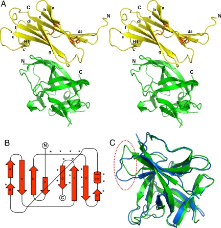
Human interleukin-18 (hIL-18) is a cytokine that plays an important role in inflammation and host defense against microbes. Its activity is regulated in vivo by a naturally occurring antagonist, the human IL-18-binding protein (IL-18BP). Functional homologs of human IL-18BP are encoded by all orthopoxviruses, including variola virus, the causative agent of smallpox. They contribute to virulence by suppressing IL-18-mediated immune responses. Here, we describe the 2.0-A resolution crystal structure of an orthopoxvirus IL-18BP, ectromelia virus IL-18BP (ectvIL-18BP), in complex with hIL-18. The hIL-18 structure in the complex shows significant conformational change at the binding interface compared with the structure of ligand-free hIL-18, indicating that the binding is mediated by an induced-fit mechanism. EctvIL-18BP adopts a canonical Ig fold and interacts via one edge of its beta-sandwich with 3 cavities on the hIL-18 surface through extensive hydrophobic and hydrogen bonding interactions. Most of the ectvIL-18BP residues that participate in these interactions are conserved in both human and viral homologs, explaining their functional equivalence despite limited sequence homology. EctvIL-18BP blocks a putative receptor-binding site on IL-18, thus preventing IL-18 from engaging its receptor. Our structure provides insights into how IL-18BPs modulate hIL-18 activity. The revealed binding interface provides the basis for rational design of inhibitors against orthopoxvirus IL-18BP (for treating orthopoxvirus infection) or hIL-18 (for treating certain inflammatory and autoimmune diseases).


Conflict of interest statement

The authors declare no conflict of interest.

Figures

Fig. 1.
Fig. 1.
Overall structure of ectvIL-18BP and hIL-18 complex. (A) Stereoview of the complex. EctvIL-18BP (yellow) sits atop the hIL-18 molecule (green), which adopts a β-trefoil structure. The 2 disulfide bonds are shown as orange sticks. The amino and carboxyl termini of the protein polypeptides are indicated as N and C, respectively. EctvIL-18BP adopts a canonical Ig fold whose secondary structures are labeled. (B) Topology diagram of ectvIL-18BP. Asterisks indicate the positions of residues interacting with hIL-18. (C) Superimposition of the crystal structure of hIL-18 (green) with the ligand-free NMR structure of hIL-18 (blue; PDB ID code 1J0S). Significant conformational changes were observed at the loop region connecting β4 and β5. Notice the loop in the crystal structure shifted by ≈6 Å from its position in ligand-free state, as indicated in a red dashed cycle.
Fig. 2.
Fig. 2.
HIL-18–ectvIL-18BP interface. (A) Three binding pockets on hIL-18 surface. (Center) EctvIL-18BP and hIL-18 complex in a top-down view as seen in Fig. 1; hIL18 is shown as surface presentation and colored gray, and ectvIL-18BP is drawn as a ribbon diagram with β-sheets colored in yellow. Binding sites A–C on the hIL-18 surface are colored red, orange, and cyan, respectively. EctvIL-18BP residues involved in binding hIL-18 are shown as sticks. (Insets) Interactions involved in the respective binding site between ectvIL-18BP and hIL-18. (B) Remodeling of hIL-18 at site A. The coloring scheme is the same as in Fig. 1C. Tyr-1, Lys-53, Ser-66, and Pro-57 are shown as sticks. Notice the drastic repositioning of the side chains of Tyr-1 and Lys-53 at the carboxyl terminus of hIL-18 upon binding ectvIL-18BP. (C and D) Results of the conformational change upon ectvIL-18BP binding, yielding a new binding cavity/pocket for Tyr-53 and Phe-67 of ectvIL-18BP (shown as sticks). The ligand-free hIL-18 is shown as a surface presentation in blue in C. The complexed hIL-18 in the current crystal structure is shown as surface model in green. Notice that the absence of Phe-67 pocket and the steric clash on Tyr-53 would occur with the ligand-free state of hIL-18. The 2 key hIL-18 residues, Lys-53 and Tyr-1 are labeled as bold italic letters.
Fig. 3.
Fig. 3.
Mechanism by which ectvIL-18BP inhibits hIL-18. EctvIL-18BP blocks the putative hIL-18Rα-binding site. Human IL-18 is represented as a surface model as viewed from the top of the β-trefoil. EctvIL-18BP-binding sites A–C on hIL-18 are colored in red, orange, and cyan, respectively. Human IL-18 residues that are potentially shared between ectvIL-18BP and hIL-18Rα are colored in purple and span the 3 ectvIL-18BP-binding sites on hIL-18.

References

    1. Okamura H, et al. Cloning of a new cytokine that induces IFN-γ production by T cells. Nature. 1995;378:88–91. - PubMed
    1. Dinarello CA. Interleukin-18. Methods. 1999;19:121–132. - PubMed
    1. Novick D, et al. Interleukin-18-binding protein: A novel modulator of the Th1 cytokine response. Immunity. 1999;10:127–136. - PubMed
    1. Xiang Y, Moss B. IL-18 binding and inhibition of interferon γ induction by human poxvirus-encoded proteins. Proc Natl Acad Sci USA. 1999;96:11537–11542. - PMC - PubMed
    1. Born TL, et al. A poxvirus protein that binds to and inactivates IL-18 and inhibits NK cell response. J Immunol. 2000;164:3246–3254. - PubMed

Publication types

MeSH terms

Substances

Associated data

LinkOut - more resources